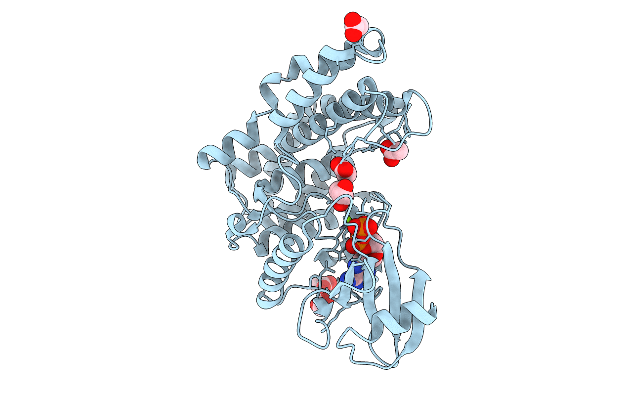
4WH2 image
Entry Detail
PDB ID:
4WH2
Keywords:
Title:
N-acetylhexosamine 1-kinase in complex with ADP
Biological Source:
PDB Version:
Deposition Date:
2014-09-19
Release Date:
2015-02-18
Method Details:
Experimental Method:
Resolution:
1.85 Å
R-Value Free:
0.24
R-Value Work:
0.19
R-Value Observed:
0.19
Space Group:
P 21 21 21
Macromolecular Entities
Polymer Type:polypeptide(L)
Description:N-acetylhexosamine 1-kinase
Mutations:D7V
Chain IDs:A
Chain Length:367
Number of Molecules:1
Biological Source:Bifidobacterium longum subsp. longum JCM 1217
Primary Citation
Open-close structural change upon ligand binding and two magnesium ions required for the catalysis of N-acetylhexosamine 1-kinase
Biochim.Biophys.Acta 1854 333 340 (2015)
PMID: 25644306 DOI: 10.1016/j.bbapap.2015.01.011

Abstact

Infant gut-associated bifidobacteria possess a metabolic pathway to utilize lacto-N-biose (Gal-β1,3-GlcNAc) and galacto-N-biose (Gal-β1,3-GalNAc) from human milk and glycoconjugates specifically. In this pathway, N-acetylhexosamine 1-kinase (NahK) catalyzes the phosphorylation of GlcNAc or GalNAc at the anomeric C1 position with ATP. Crystal structures of NahK have only been determined in the closed state. In this study, we determined open state structures of NahK in three different forms (apo, ADP complex, and ATP complex). A comparison of the open and closed state structures revealed an induced fit structural change defined by two rigid domains. ATP binds to the small N-terminal domain, and binding of the N-acetylhexosamine substrate to the large C-terminal domain induces a closing conformational change with a rotation angle of 16°. In the nucleotide binding site, two magnesium ions bridging the α-γ and β-γ phosphates were identified. A mutational analysis indicated that a residue coordinating both of the two magnesium ions (Asp228) is essential for catalysis. The involvement of two magnesium ions in the catalytic machinery is structurally similar to the catalytic structures of protein kinases and aminoglycoside phosphotransferases, but distinct from the structures of other anomeric kinases or sugar 6-kinases. These findings help to elucidate the possible evolutionary adaptation of substrate specificities and induced fit mechanism.

Legend

Protein

Chemical

Disease

Primary Citation of related structures