Deposition Date
2014-09-10
Release Date
2015-02-04
Last Version Date
2024-01-10
Entry Detail
PDB ID:
4WEI
Keywords:
Title:
Crystal structure of the F4 fimbrial adhesin FaeG in complex with lactose
Biological Source:
Source Organism(s):
Escherichia coli (Taxon ID: 562)
Expression System(s):
Method Details:
Experimental Method:
Resolution:
2.30 Å
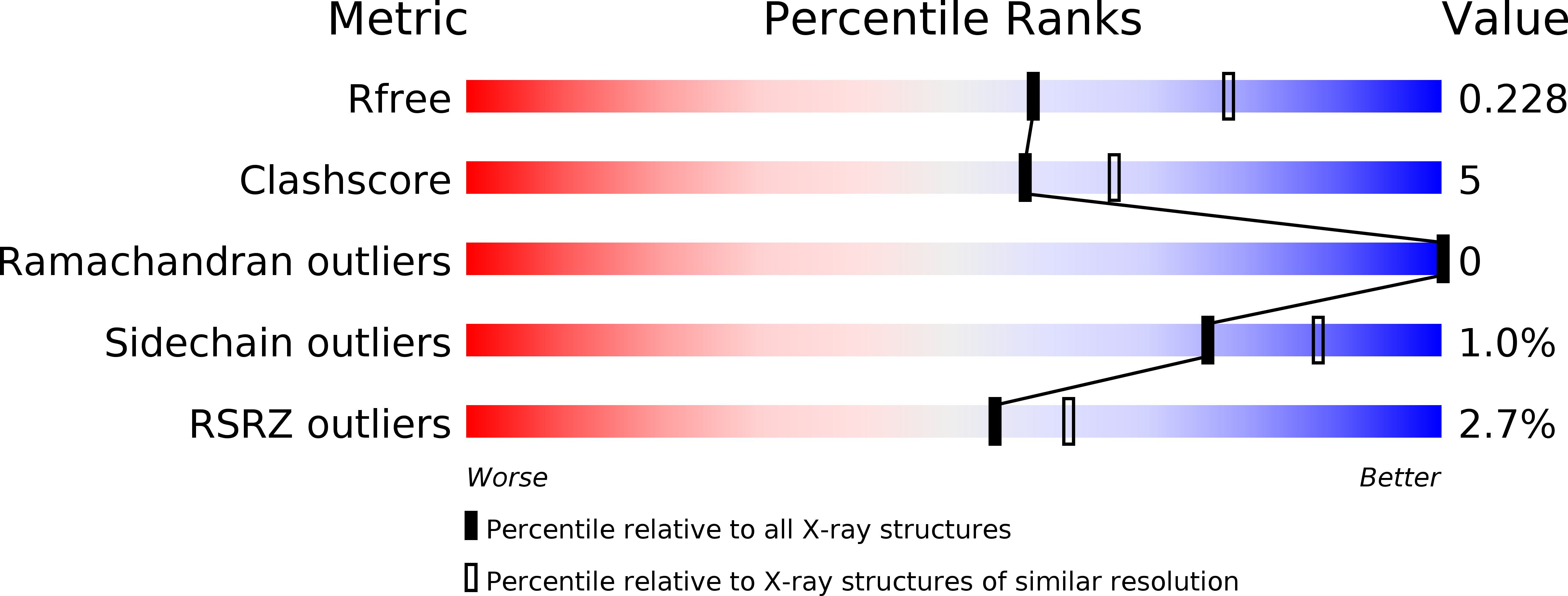
R-Value Free:
0.23
R-Value Work:
0.19
R-Value Observed:
0.19
Space Group:
P 41 21 2