
Deposition Date
2014-09-05
Release Date
2014-09-24
Last Version Date
2023-09-27
Entry Detail
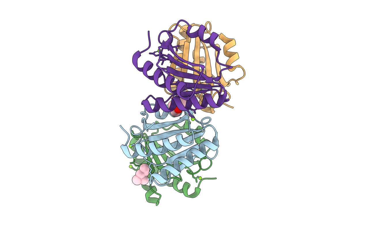
PDB ID:
4WCW
Keywords:
Title:
Ribosomal silencing factor during starvation or stationary phase (RsfS) from Mycobacterium tuberculosis
Biological Source:
Source Organism(s):
Mycobacterium tuberculosis (Taxon ID: 83332)
Expression System(s):
Method Details:
Experimental Method:
Resolution:
2.10 Å
R-Value Free:
0.25
R-Value Work:
0.21
R-Value Observed:
0.21
Space Group:
P 1
