Deposition Date
2014-09-04
Release Date
2014-11-12
Last Version Date
2023-09-27
Entry Detail
Biological Source:
Source Organism(s):
Ammonifex degensii (Taxon ID: 429009)
Expression System(s):
Method Details:
Experimental Method:
Resolution:
1.70 Å
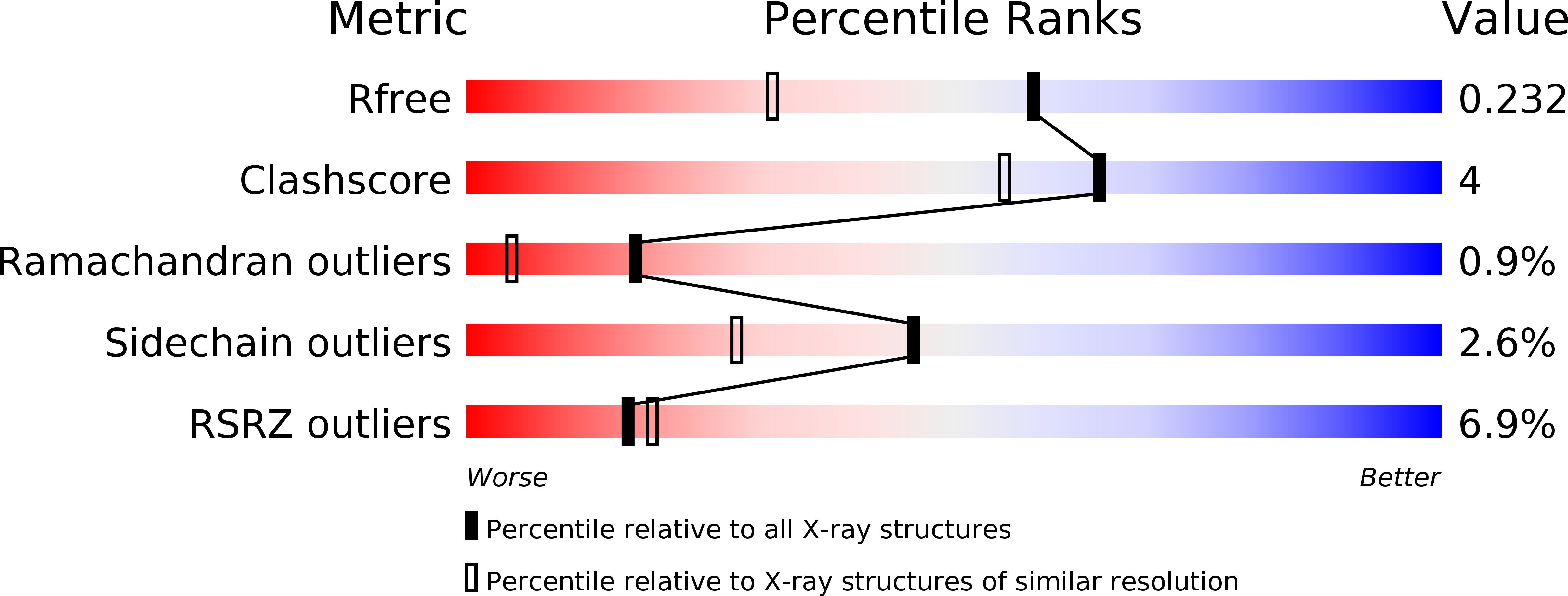
R-Value Free:
0.23
R-Value Work:
0.20
R-Value Observed:
0.20
Space Group:
I 4