4WBD image
Entry Detail
PDB ID:
4WBD
Keywords:
Title:
The crystal structure of BshC from Bacillus subtilis complexed with citrate and ADP
Biological Source:
Source Organism:
Host Organism:
PDB Version:
Deposition Date:
2014-09-03
Release Date:
2014-12-31
Method Details:
Experimental Method:
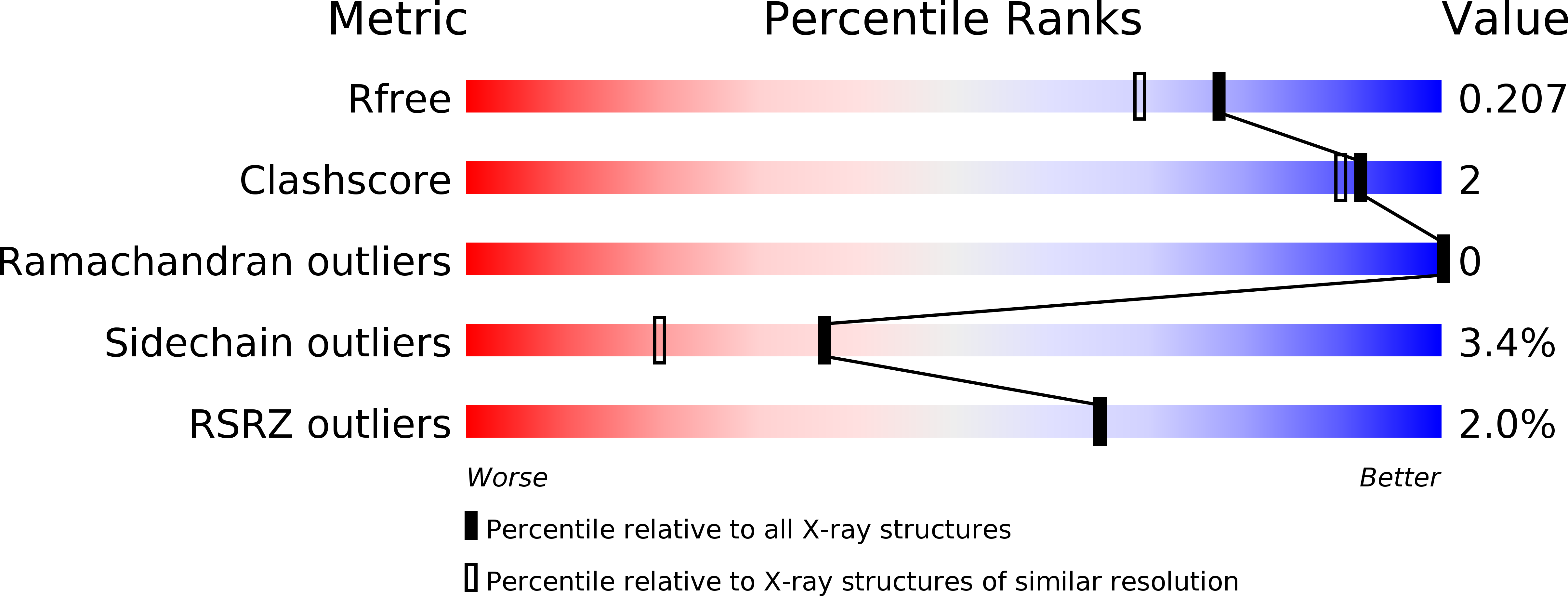
Resolution:
1.77 Å
R-Value Free:
0.19
R-Value Work:
0.16
R-Value Observed:
0.16
Space Group:
I 1 2 1
Macromolecular Entities
Polymer Type:polypeptide(L)
Description:BSHC
Chain IDs:A
Chain Length:541
Number of Molecules:1
Biological Source:Bacillus subtilis
Primary Citation
X-ray Crystallographic Structure of BshC, a Unique Enzyme Involved in Bacillithiol Biosynthesis.
Biochemistry 54 100 103 (2015)
PMID: 25496067 DOI: 10.1021/bi501394q

Abstact

Bacillithiol is produced by many Gram-positive bacteria via a pathway utilizing the enzymes BshA, BshB, and BshC. Here we report the 1.77 Å resolution crystal structure of BshC, the putative cysteine ligase in bacillithiol production. The structure reveals that BshC contains a core Rossmann fold with connecting peptide motifs (CP1 and CP2) and a unique α-helical coiled-coil domain that facilitates dimerization. The model contains citrate and glycerol in the canonical active site and ADP in a second binding pocket. The overall structure and bound ligands give insight into the function of this unique enzyme.

Legend

Protein

Chemical

Disease

Primary Citation of related structures