4W7K image
Entry Detail
PDB ID:
4W7K
Keywords:
Title:
CRYSTAL STRUCTURE OF A DECOLORIZING PEROXIDASE (DYP) FROM AURICULARIA AURICULA-JUDAE. Y147S MUTANT
Biological Source:
Host Organism:
PDB Version:
Deposition Date:
2014-08-22
Release Date:
2015-03-11
Method Details:
Experimental Method:
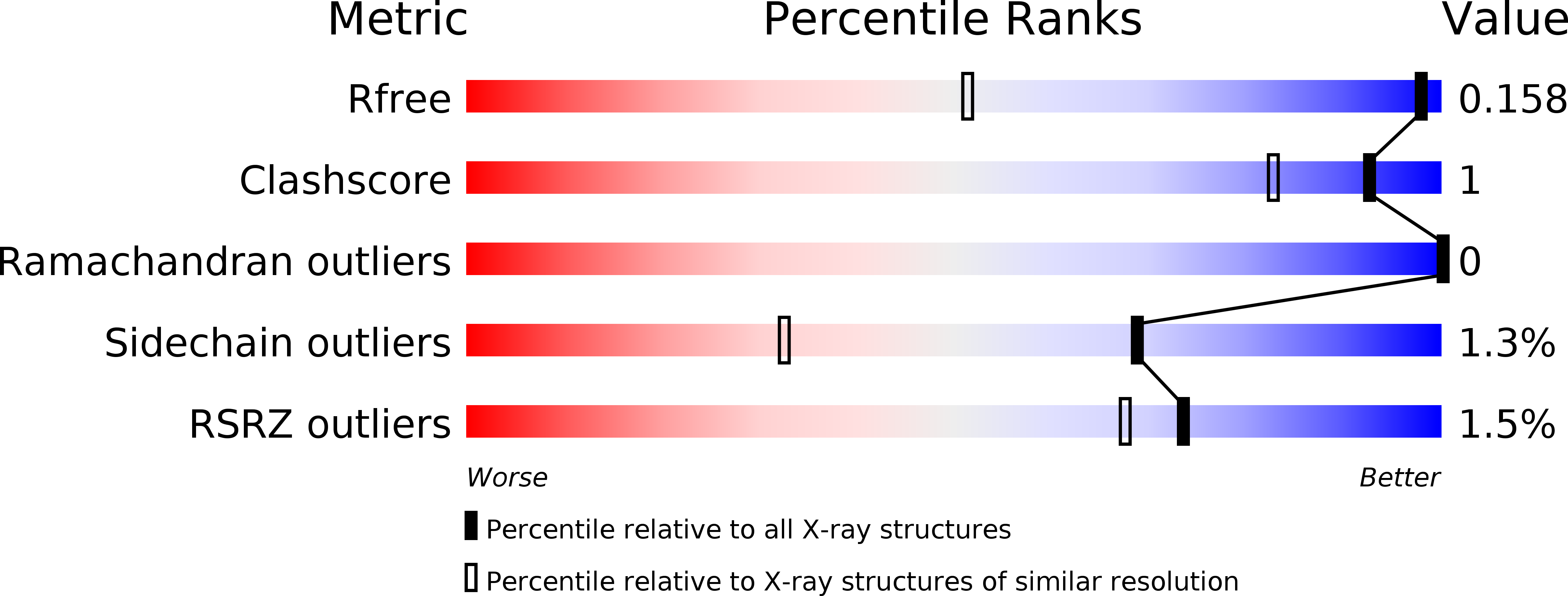
Resolution:
1.05 Å
R-Value Free:
0.15
R-Value Work:
0.13
R-Value Observed:
0.13
Space Group:
C 1 2 1
Macromolecular Entities
Polymer Type:polypeptide(L)
Description:Dye-decolorizing peroxidase
Mutations:yes
Chain IDs:A, B
Chain Length:449
Number of Molecules:2
Biological Source:Auricularia auricula-judae
Ligand Molecules
Primary Citation
Catalytic surface radical in dye-decolorizing peroxidase: a computational, spectroscopic and site-directed mutagenesis study.
Biochem.J. 466 253 262 (2015)
PMID: 25495127 DOI: 10.1042/BJ20141211

Abstact

Dye-decolorizing peroxidase (DyP) of Auricularia auricula-judae has been expressed in Escherichia coli as a representative of a new DyP family, and subjected to mutagenic, spectroscopic, crystallographic and computational studies. The crystal structure of DyP shows a buried haem cofactor, and surface tryptophan and tyrosine residues potentially involved in long-range electron transfer from bulky dyes. Simulations using PELE (Protein Energy Landscape Exploration) software provided several binding-energy optima for the anthraquinone-type RB19 (Reactive Blue 19) near the above aromatic residues and the haem access-channel. Subsequent QM/MM (quantum mechanics/molecular mechanics) calculations showed a higher tendency of Trp-377 than other exposed haem-neighbouring residues to harbour a catalytic protein radical, and identified the electron-transfer pathway. The existence of such a radical in H₂O₂-activated DyP was shown by low-temperature EPR, being identified as a mixed tryptophanyl/tyrosyl radical in multifrequency experiments. The signal was dominated by the Trp-377 neutral radical contribution, which disappeared in the W377S variant, and included a tyrosyl contribution assigned to Tyr-337 after analysing the W377S spectra. Kinetics of substrate oxidation by DyP suggests the existence of high- and low-turnover sites. The high-turnover site for oxidation of RB19 (k(cat) > 200 s⁻¹) and other DyP substrates was assigned to Trp-377 since it was absent from the W377S variant. The low-turnover site/s (RB19 k(cat) ~20 s⁻¹) could correspond to the haem access-channel, since activity was decreased when the haem channel was occluded by the G169L mutation. If a tyrosine residue is also involved, it will be different from Tyr-337 since all activities are largely unaffected in the Y337S variant.

Legend

Protein

Chemical

Disease

Primary Citation of related structures