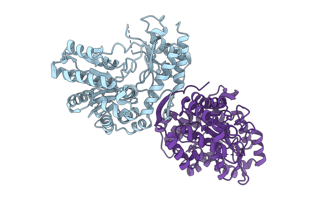
4W4H image
Entry Detail
PDB ID:
4W4H
Keywords:
Title:
Escherichia coli tryptophanase in holo form
Biological Source:
Source Organism:
Host Organism:
PDB Version:
Deposition Date:
2014-08-14
Release Date:
2014-12-17
Method Details:
Experimental Method:
Resolution:
2.89 Å
R-Value Free:
0.27
R-Value Work:
0.22
R-Value Observed:
0.22
Space Group:
P 43 21 2
Macromolecular Entities
Polymer Type:polypeptide(L)
Description:Tryptophanase
Chain IDs:A, B
Chain Length:467
Number of Molecules:2
Biological Source:Escherichia coli
Modified Residue
Compound ID Chain ID Parent Comp ID Details 2D Image
LLP A LYS modified residue
Primary Citation
Structures of Escherichia coli tryptophanase in holo and `semi-holo' forms.
Acta Crystallogr.,Sect.F 71 286 290 (2015)
PMID: 25760702 DOI: 10.1107/S2053230X15000850

Abstact

Two crystal forms of Escherichia coli tryptophanase (tryptophan indole-lyase, Trpase) were obtained under the same crystallization conditions. Both forms belonged to the same space group P43212 but had slightly different unit-cell parameters. The holo crystal form, with pyridoxal phosphate (PLP) bound to Lys270 of both polypeptide chains in the asymmetric unit, diffracted to 2.9 Å resolution. The second crystal form diffracted to 3.2 Å resolution. Of the two subunits in the asymmetric unit, one was found in the holo form, while the other appeared to be in the apo form in a wide-open conformation with two sulfate ions bound in the vicinity of the active site. The conformation of all holo subunits is the same in both crystal forms. The structures suggest that Trpase is flexible in the apo form. Its conformation partially closes upon binding of PLP. The closed conformation might correspond to the enzyme in its active state with both cofactor and substrate bound in a similar way as in tyrosine phenol-lyase.

Legend

Protein

Chemical

Disease

Primary Citation of related structures