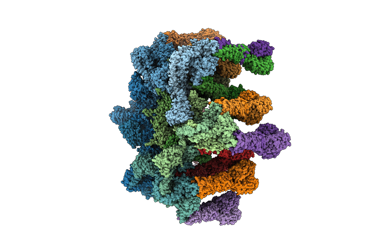
4V96 image
Entry Detail
PDB ID:
4V96
Keywords:
Title:
The structure of a 1.8 MDa viral genome injection device suggests alternative infection mechanisms
Biological Source:
Source Organism:
Host Organism:
PDB Version:
Deposition Date:
2012-02-01
Release Date:
2014-07-09
Method Details:
Experimental Method:
Resolution:
3.80 Å
R-Value Free:
0.26
R-Value Work:
0.23
R-Value Observed:
0.23
Space Group:
P 1 21 1
Macromolecular Entities
Polymer Type:polypeptide(L)
Description:ORF48
Chain IDs:A (auth: AA), B (auth: AB), C (auth: AC), D (auth: AD), E (auth: AE), F (auth: AF), G (auth: AG), H (auth: AH), I (auth: AI), J (auth: AJ), K (auth: AK), L (auth: AL), M (auth: AM), N (auth: AN), O (auth: AO), P (auth: AP), Q (auth: AQ), R (auth: AR)
Chain Length:299
Number of Molecules:18
Biological Source:Lactococcus phage TP901-1
Polymer Type:polypeptide(L)
Description:ORF46
Chain IDs:S (auth: AS), T (auth: AT), U (auth: AU), V (auth: AV), W (auth: AW), X (auth: AX)
Chain Length:253
Number of Molecules:6
Biological Source:Lactococcus phage TP901-1
Polymer Type:polypeptide(L)
Description:BPP
Chain IDs:Y (auth: BA), Z (auth: BB), AA (auth: BC), BA (auth: BD), CA (auth: BE), DA (auth: BF), EA (auth: BG), FA (auth: BH), GA (auth: BI), HA (auth: BJ), IA (auth: BK), JA (auth: BL), KA (auth: BM), LA (auth: BN), MA (auth: BO), NA (auth: BP), OA (auth: BQ), PA (auth: BR), QA (auth: BS), RA (auth: BT), SA (auth: BU), TA (auth: BV), UA (auth: BW), VA (auth: BX), WA (auth: BY), XA (auth: BZ), YA (auth: Ba), ZA (auth: Bb), AB (auth: Bc), BB (auth: Bd), CB (auth: Be), DB (auth: Bf), EB (auth: Bg), FB (auth: Bh), GB (auth: Bi), HB (auth: Bj), IB (auth: Bk), JB (auth: Bl), KB (auth: Bm), LB (auth: Bn), MB (auth: Bo), NB (auth: Bp), OB (auth: Bq), PB (auth: Br), QB (auth: Bs), RB (auth: Bt), SB (auth: Bu), TB (auth: Bv), UB (auth: Bw), VB (auth: Bx), WB (auth: By), XB (auth: Bz), YB (auth: B1), ZB (auth: B2)
Chain Length:173
Number of Molecules:54
Biological Source:Lactococcus phage TP901-1
Ligand Molecules
Primary Citation
Structure of the phage TP901-1 1.8 MDa baseplate suggests an alternative host adhesion mechanism.
Proc.Natl.Acad.Sci.USA 109 8954 8958 (2012)
PMID: 22611190 DOI: 10.1073/pnas.1200966109

Abstact

Phages of the Caudovirales order possess a tail that recognizes the host and ensures genome delivery upon infection. The X-ray structure of the approximately 1.8 MDa host adsorption device (baseplate) from the lactococcal phage TP901-1 shows that the receptor-binding proteins are pointing in the direction of the host, suggesting that this organelle is in a conformation ready for host adhesion. This result is in marked contrast with the lactococcal phage p2 situation, whose baseplate is known to undergo huge conformational changes in the presence of Ca(2+) to reach its active state. In vivo infection experiments confirmed these structural observations by demonstrating that Ca(2+) ions are required for host adhesion among p2-like phages (936-species) but have no influence on TP901-1-like phages (P335-species). These data suggest that these two families rely on diverse adhesion strategies which may lead to different signaling for genome release.

Legend

Protein

Chemical

Disease

Primary Citation of related structures