4V6B image
Deposition Date 2009-06-19
Release Date 2014-07-09
Last Version Date 2023-09-20
Entry Detail
PDB ID:
4V6B
Keywords:
Title:
Crystal structure of human ferritin Phe167SerfsX26 mutant.
Biological Source:
Source Organism:
Homo sapiens (Taxon ID: 9606)
Host Organism:
Method Details:
Experimental Method:
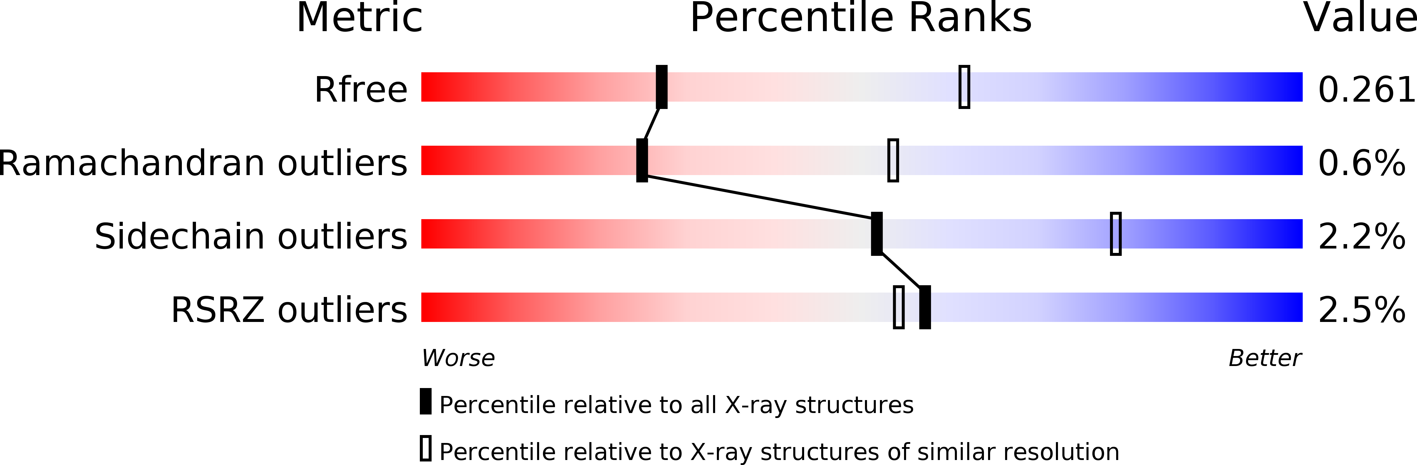
Resolution:
2.85 Å
R-Value Free:
0.29
R-Value Work:
0.24
Space Group:
P 1
Macromolecular Entities
Structures with similar UniProt ID
Protein Blast
Polymer Type:polypeptide(L)
Molecule:Ferritin
Gene (Uniprot):FTL
Mutagens:Phe167SerfsX26
Chain IDs:A (auth: AA), B (auth: Aa), C (auth: AB), D (auth: Ab), E (auth: AC), F (auth: Ac), G (auth: AD), H (auth: Ad), I (auth: AE), J (auth: Ae), K (auth: AF), L (auth: Af), M (auth: AG), N (auth: Ag), O (auth: AH), P (auth: Ah), Q (auth: AI), R (auth: Ai), S (auth: AJ), T (auth: Aj), U (auth: AK), V (auth: Ak), W (auth: AL), X (auth: Al), Y (auth: AM), Z (auth: Am), AA (auth: AN), BA (auth: An), CA (auth: AO), DA (auth: Ao), EA (auth: AP), FA (auth: Ap), GA (auth: AQ), HA (auth: Aq), IA (auth: AR), JA (auth: Ar), KA (auth: AS), LA (auth: As), MA (auth: AT), NA (auth: At), OA (auth: AU), PA (auth: Au), QA (auth: AV), RA (auth: Av), SA (auth: AW), TA (auth: Aw), UA (auth: AX), VA (auth: Ax), WA (auth: BA), XA (auth: Ba), YA (auth: BB), ZA (auth: Bb), AB (auth: BC), BB (auth: Bc), CB (auth: BD), DB (auth: Bd), EB (auth: BE), FB (auth: Be), GB (auth: BF), HB (auth: Bf), IB (auth: BG), JB (auth: Bg), KB (auth: BH), LB (auth: Bh), MB (auth: BI), NB (auth: Bi), OB (auth: BJ), PB (auth: Bj), QB (auth: BK), RB (auth: Bk), SB (auth: BL), TB (auth: Bl), UB (auth: BM), VB (auth: Bm), WB (auth: BN), XB (auth: Bn), YB (auth: BO), ZB (auth: Bo), AC (auth: BP), BC (auth: Bp), CC (auth: BQ), DC (auth: Bq), EC (auth: BR), FC (auth: Br), GC (auth: BS), HC (auth: Bs), IC (auth: BT), JC (auth: Bt), KC (auth: BU), LC (auth: Bu), MC (auth: BV), NC (auth: Bv), OC (auth: BW), PC (auth: Bw), QC (auth: BX), RC (auth: Bx), SC (auth: CA), TC (auth: Ca), UC (auth: CB), VC (auth: Cb), WC (auth: CC), XC (auth: Cc), YC (auth: CD), ZC (auth: Cd), AD (auth: CE), BD (auth: Ce), CD (auth: CF), DD (auth: Cf), ED (auth: CG), FD (auth: Cg), GD (auth: CH), HD (auth: Ch), ID (auth: CI), JD (auth: Ci), KD (auth: CJ), LD (auth: Cj), MD (auth: CK), ND (auth: Ck), OD (auth: CL), PD (auth: Cl), QD (auth: CM), RD (auth: Cm), SD (auth: CN), TD (auth: Cn), UD (auth: CO), VD (auth: Co), WD (auth: CP), XD (auth: Cp), YD (auth: CQ), ZD (auth: Cq), AE (auth: CR), BE (auth: Cr), CE (auth: CS), DE (auth: Cs), EE (auth: CT), FE (auth: Ct), GE (auth: CU), HE (auth: Cu), IE (auth: CV), JE (auth: Cv), KE (auth: CW), LE (auth: Cw), ME (auth: CX), NE (auth: Cx)
Chain Length:192
Number of Molecules:144
Biological Source:Homo sapiens
Ligand Molecules
Primary Citation
Unraveling of the E-helices and disruption of 4-fold pores are associated with iron mishandling in a mutant ferritin causing neurodegeneration
J.Biol.Chem. 285 1950 1956 (2010)
PMID: 19923220 DOI: 10.1074/jbc.M109.042986

Abstact

Mutations in the coding sequence of the ferritin light chain (FTL) gene cause a neurodegenerative disease known as neuroferritinopathy or hereditary ferritinopathy, which is characterized by the presence of intracellular inclusion bodies containing the mutant FTL polypeptide and by abnormal accumulation of iron in the brain. Here, we describe the x-ray crystallographic structure and report functional studies of ferritin homopolymers formed from the mutant FTL polypeptide p.Phe167SerfsX26, which has a C terminus that is altered in amino acid sequence and length. The structure was determined and refined to 2.85 A resolution and was very similar to the wild type between residues Ile-5 and Arg-154. However, instead of the E-helices normally present in wild type ferritin, the C-terminal sequences of all 24 mutant subunits showed substantial amounts of disorder, leading to multiple C-terminal polypeptide conformations and a large disruption of the normally tiny 4-fold axis pores. Functional studies underscored the importance of the mutant C-terminal sequence in iron-induced precipitation and revealed iron mishandling by soluble mutant FTL homopolymers in that only wild type incorporated iron when in direct competition in solution with mutant ferritin. Even without competition, the amount of iron incorporation over the first few minutes differed severalfold. Our data suggest that disruption at the 4-fold pores may lead to direct iron mishandling through attenuated iron incorporation by the soluble form of mutant ferritin and that the disordered C-terminal polypeptides may play a major role in iron-induced precipitation and formation of ferritin inclusion bodies in hereditary ferritinopathy.

Legend

Protein

Chemical

Disease

Primary Citation of related structures