4V5I image
Entry Detail
PDB ID:
4V5I
Keywords:
Title:
Structure of the Phage P2 Baseplate in its Activated Conformation with Ca
Biological Source:
Source Organism:
Host Organism:
PDB Version:
Deposition Date:
2010-02-05
Release Date:
2014-07-09
Method Details:
Experimental Method:
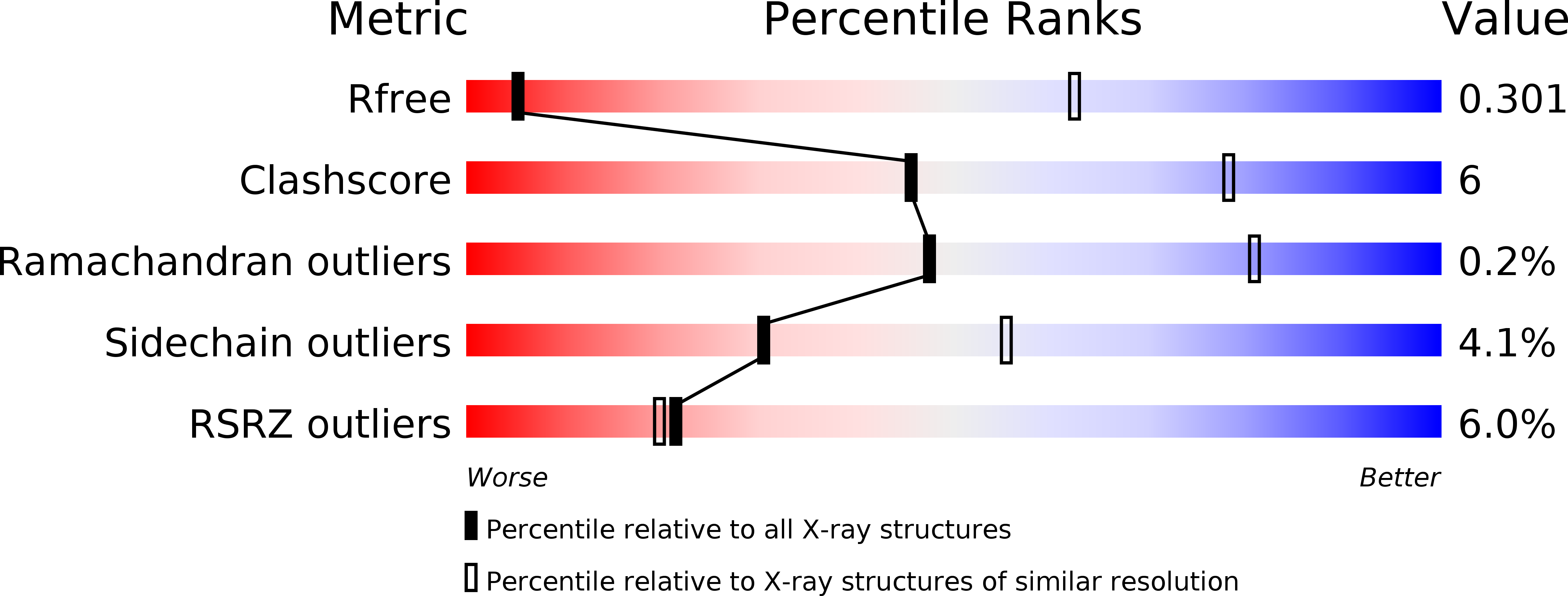
Resolution:
5.46 Å
R-Value Free:
0.29
R-Value Work:
0.29
R-Value Observed:
0.29
Space Group:
P 1 21 1
Macromolecular Entities
Polymer Type:polypeptide(L)
Description:ORF16
Chain IDs:A (auth: A0), Z (auth: AY), AA (auth: AZ), BA (auth: B0), AB (auth: BY), BB (auth: BZ)
Chain Length:372
Number of Molecules:6
Biological Source:LACTOCOCCUS PHAGE P2
Polymer Type:polypeptide(L)
Description:PUTATIVE RECEPTOR BINDING PROTEIN
Chain IDs:B (auth: AA), C (auth: AB), D (auth: AC), E (auth: AD), F (auth: AE), G (auth: AF), H (auth: AG), I (auth: AH), J (auth: AI), K (auth: AJ), L (auth: AK), M (auth: AL), N (auth: AM), O (auth: AN), P (auth: AO), Q (auth: AP), R (auth: AQ), S (auth: AR), CA (auth: BA), DA (auth: BB), EA (auth: BC), FA (auth: BD), GA (auth: BE), HA (auth: BF), IA (auth: BG), JA (auth: BH), KA (auth: BI), LA (auth: BJ), MA (auth: BK), NA (auth: BL), OA (auth: BM), PA (auth: BN), QA (auth: BO), RA (auth: BP), SA (auth: BQ), TA (auth: BR)
Chain Length:263
Number of Molecules:36
Biological Source:LACTOCOCCUS PHAGE P2
Polymer Type:polypeptide(L)
Description:ORF15
Chain IDs:T (auth: AS), U (auth: AT), V (auth: AU), W (auth: AV), X (auth: AW), Y (auth: AX), UA (auth: BS), VA (auth: BT), WA (auth: BU), XA (auth: BV), YA (auth: BW), ZA (auth: BX)
Chain Length:298
Number of Molecules:12
Biological Source:LACTOCOCCUS PHAGE P2
Ligand Molecules
Primary Citation
Structure of Lactococcal Phage P2 Baseplate and its Mechanism of Activation.
Proc.Natl.Acad.Sci.USA 107 6852 ? (2010)
PMID: 20351260 DOI: 10.1073/PNAS.1000232107

Abstact

Siphoviridae is the most abundant viral family on earth which infects bacteria as well as archaea. All known siphophages infecting gram+ Lactococcus lactis possess a baseplate at the tip of their tail involved in host recognition and attachment. Here, we report analysis of the p2 phage baseplate structure by X-ray crystallography and electron microscopy and propose a mechanism for the baseplate activation during attachment to the host cell. This approximately 1 MDa, Escherichia coli-expressed baseplate is composed of three protein species, including six trimers of the receptor-binding protein (RBP). RBPs host-recognition domains point upwards, towards the capsid, in agreement with the electron-microscopy map of the free virion. In the presence of Ca(2+), a cation mandatory for infection, the RBPs rotated 200 degrees downwards, presenting their binding sites to the host, and a channel opens at the bottom of the baseplate for DNA passage. These conformational changes reveal a novel siphophage activation and host-recognition mechanism leading ultimately to DNA ejection.

Legend

Protein

Chemical

Disease

Primary Citation of related structures