4V4C image
Entry Detail
PDB ID:
4V4C
Keywords:
Title:
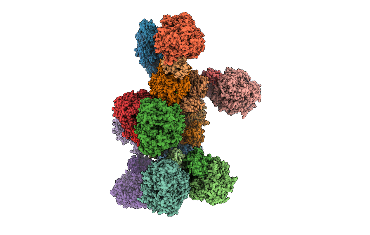
Crystal Structure of Pyrogallol-Phloroglucinol Transhydroxylase from Pelobacter acidigallici
Biological Source:
Source Organism:
PDB Version:
Deposition Date:
2004-06-02
Release Date:
2014-07-09
Method Details:
Experimental Method:
Resolution:
2.35 Å
R-Value Free:
0.25
R-Value Work:
0.19
R-Value Observed:
0.19
Space Group:
P 1
Macromolecular Entities
Polymer Type:polypeptide(L)
Description:Pyrogallol hydroxytransferase large subunit
Chain IDs:A, C, E, G, I, K, M, O, Q, S, U, W
Chain Length:875
Number of Molecules:12
Biological Source:Pelobacter acidigallici
Polymer Type:polypeptide(L)
Description:Pyrogallol hydroxytransferase small subunit
Chain IDs:B, D, F, H, J, L, N, P, R, T, V, X
Chain Length:274
Number of Molecules:12
Biological Source:Pelobacter acidigallici
Primary Citation
Crystal structure of pyrogallol-phloroglucinol transhydroxylase, an Mo enzyme capable of intermolecular hydroxyl transfer between phenols
PROC.NATL.ACAD.SCI.USA 101 11571 11576 (2004)
PMID: 15284442 DOI: 10.1073/pnas.0404378101

Abstact

The Mo enzyme transhydroxylase from the anaerobic microorganism Pelobacter acidigallici catalyzes the conversion of pyrogallol to phloroglucinol. Such trihydroxybenzenes and their derivatives represent important building blocks of plant polymers. None of the transferred hydroxyl groups originates from water during transhydroxylation; instead a cosubstrate, such as 1,2,3,5-tetrahydroxybenzene, is used in a reaction without apparent electron transfer. Here, we report on the crystal structure of the enzyme in the reduced Mo(IV) state, which we solved by single anomalous-diffraction technique. It represents the largest structure (1,149 amino acid residues per molecule, 12 independent molecules per unit cell), which has been solved so far by single anomalous-diffraction technique. Tranhydroxylase is a heterodimer, with the active Mo-molybdopterin guanine dinucleotide (MGD)(2) site in the alpha-subunit, and three [4Fe-4S] centers in the beta-subunit. The latter subunit carries a seven-stranded, mainly antiparallel beta-barrel domain. We propose a scheme for the transhydroxylation reaction based on 3D structures of complexes of the enzyme with various polyphenols serving either as substrate or inhibitor.

Legend

Protein

Chemical

Disease

Primary Citation of related structures