
Deposition Date
2014-10-13
Release Date
2015-12-09
Last Version Date
2024-10-23
Entry Detail
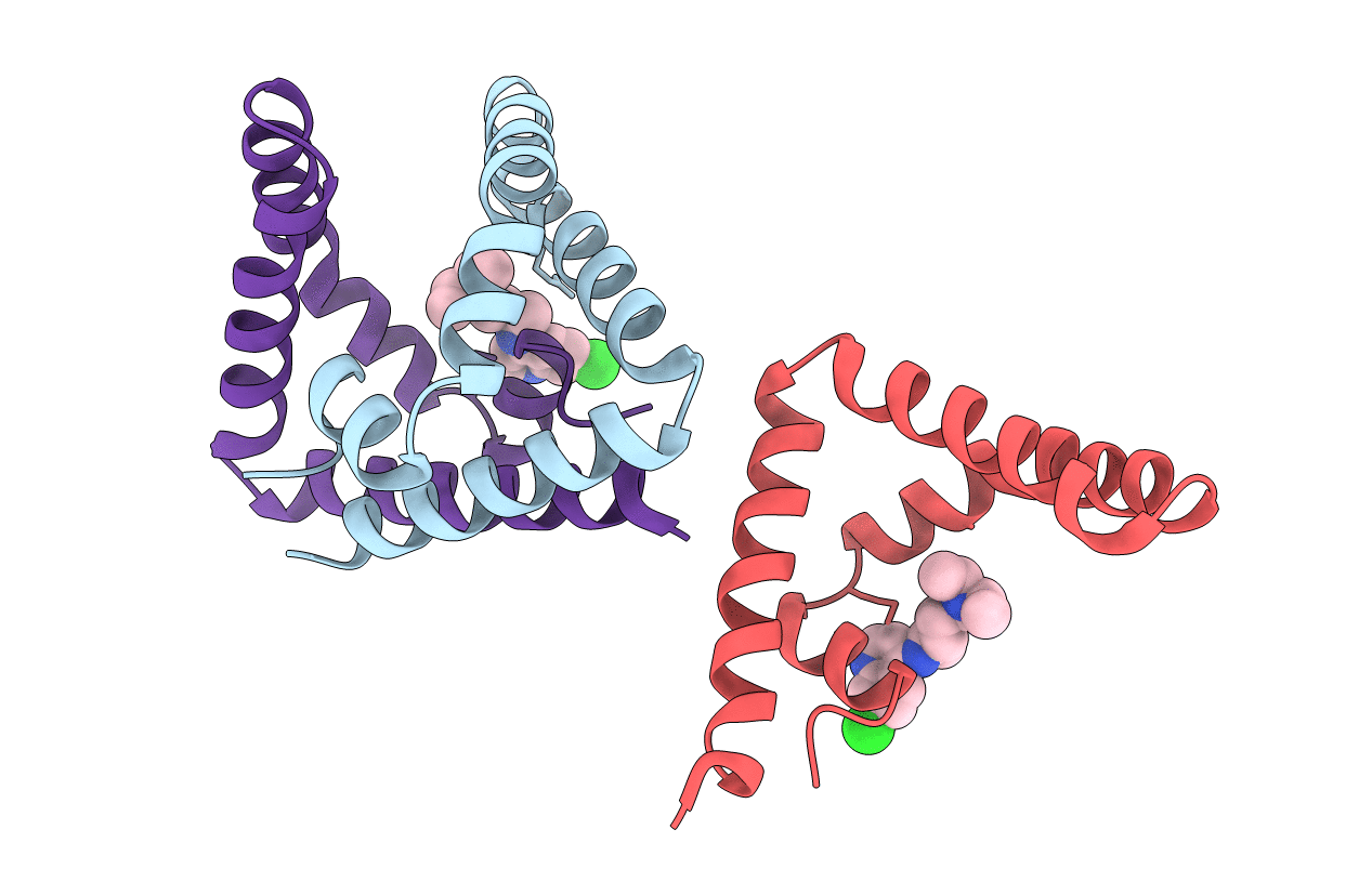
PDB ID:
4V2O
Keywords:
Title:
Structure of saposin B in complex with chloroquine
Biological Source:
Source Organism(s):
HOMO SAPIENS (Taxon ID: 9606)
Expression System(s):
Method Details:
Experimental Method:
Resolution:
2.13 Å
R-Value Free:
0.25
R-Value Work:
0.22
R-Value Observed:
0.23
Space Group:
P 32 2 1
