4V1Z image
Deposition Date 2014-10-04
Release Date 2015-01-14
Last Version Date 2024-10-16
Entry Detail
PDB ID:
4V1Z
Keywords:
Title:
The 3-D structure of the cellobiohydrolase, Cel7A, from Aspergillus fumigatus
Biological Source:
Source Organism:
Host Organism:
Method Details:
Experimental Method:
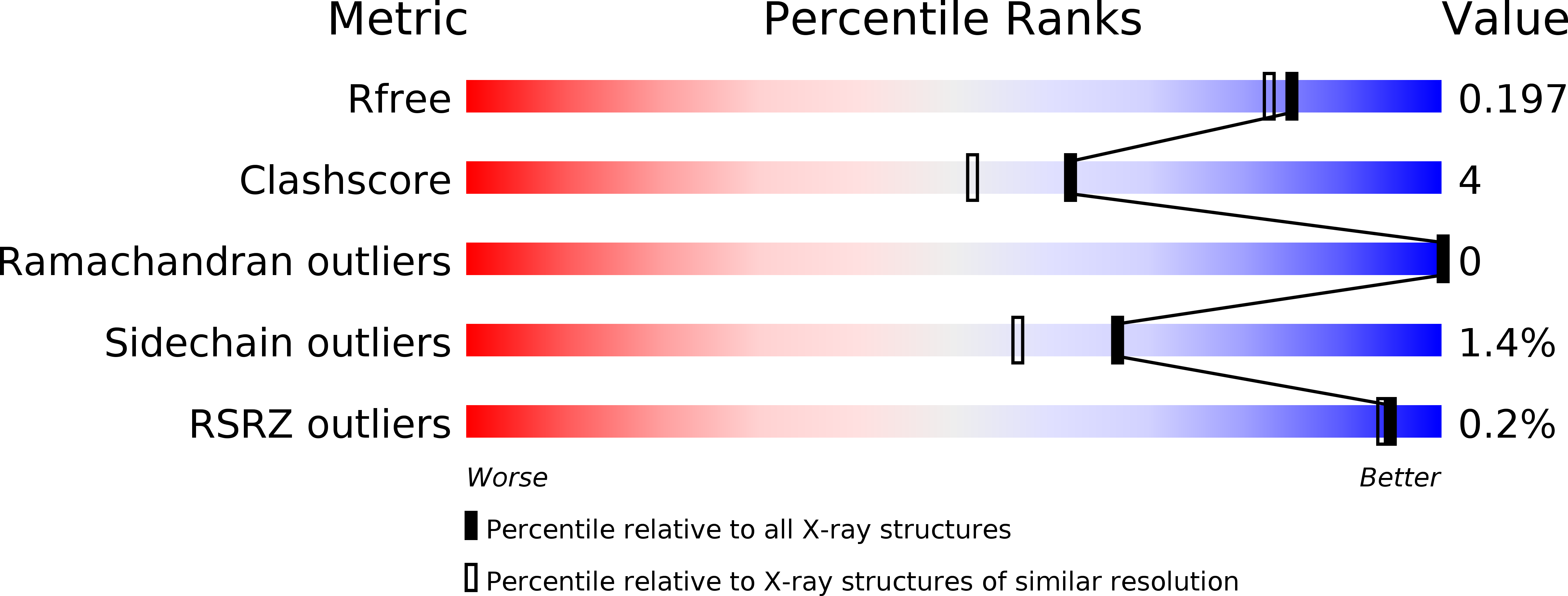
Resolution:
1.78 Å
R-Value Free:
0.18
R-Value Work:
0.14
R-Value Observed:
0.14
Space Group:
P 21 21 2
Macromolecular Entities
Structures with similar UniProt ID
Protein Blast
Polymer Type:polypeptide(L)
Molecule:CELLOBIOHYDROLASE
Gene (Uniprot):cbhB
Chain IDs:A
Chain Length:440
Number of Molecules:1
Biological Source:ASPERGILLUS FUMIGATUS
Modified Residue
Compound ID Chain ID Parent Comp ID Details 2D Image
ASN A ASN GLYCOSYLATION SITE
PCA A GLU PYROGLUTAMIC ACID
Primary Citation
The Three-Dimensional Structure of the Cellobiohydrolase Cel7A from Aspergillus Fumigatus at 1.5 A Resolution
Acta Crystallogr.,Sect.F 71 114 ? (2015)
PMID: 25615982 DOI: 10.1107/S2053230X14027307

Abstact

The enzymatic degradation of plant cell-wall cellulose is central to many industrial processes, including second-generation biofuel production. Key players in this deconstruction are the fungal cellobiohydrolases (CBHs), notably those from family GH7 of the carbohydrate-active enzymes (CAZY) database, which are generally known as CBHI enzymes. Here, three-dimensional structures are reported of the Aspergillus fumigatus CBHI Cel7A solved in uncomplexed and disaccharide-bound forms at resolutions of 1.8 and 1.5 Å, respectively. The product complex with a disaccharide in the +1 and +2 subsites adds to the growing three-dimensional insight into this family of industrially relevant biocatalysts.

Legend

Protein

Chemical

Disease

Primary Citation of related structures