4V0P image
Deposition Date 2014-09-17
Release Date 2014-10-01
Last Version Date 2024-01-10
Entry Detail
PDB ID:
4V0P
Keywords:
Title:
Crystal structure of the MAGE homology domain of human MAGE-A3
Biological Source:
Source Organism:
HOMO SAPIENS (Taxon ID: 9606)
Host Organism:
Method Details:
Experimental Method:
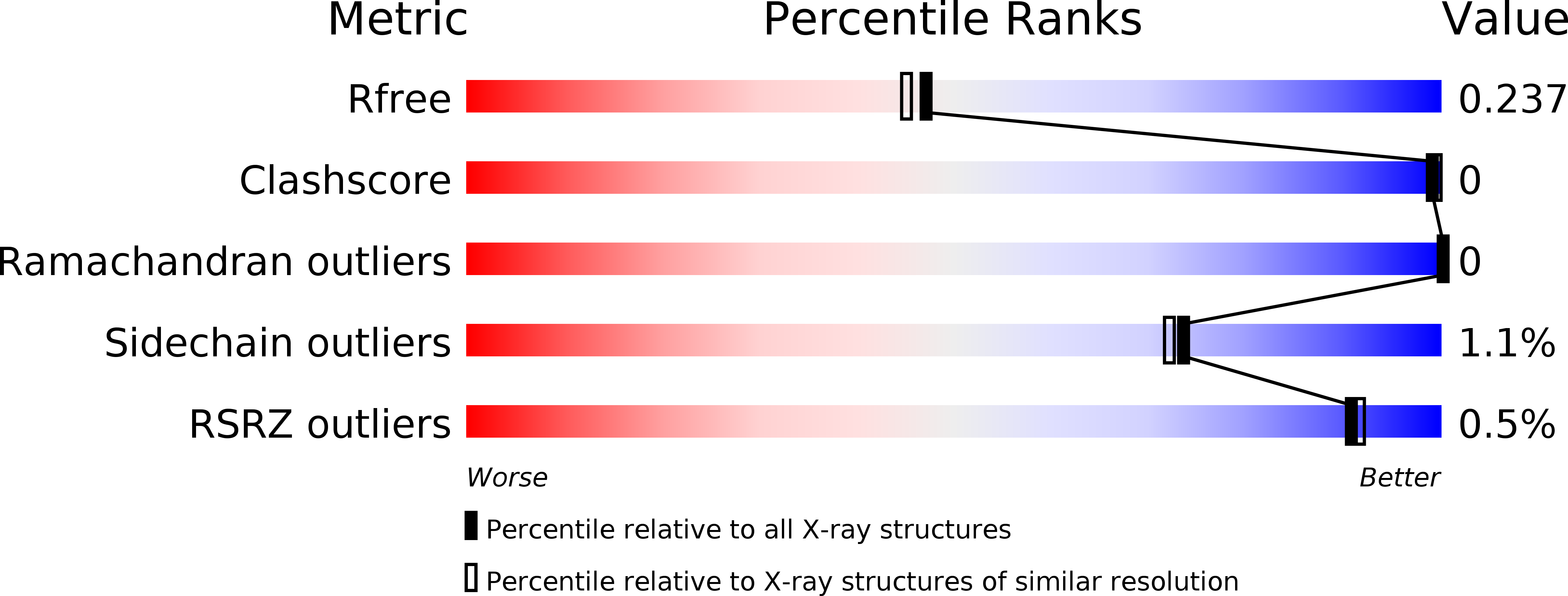
Resolution:
2.07 Å
R-Value Free:
0.23
R-Value Work:
0.20
R-Value Observed:
0.20
Space Group:
P 61 2 2
Macromolecular Entities
Polymer Type:polypeptide(L)
Molecule:MELANOMA-ASSOCIATED ANTIGEN 3
Gene (Uniprot):MAGEA3
Chain IDs:A
Chain Length:213
Number of Molecules:1
Biological Source:HOMO SAPIENS
Primary Citation
Structures of Two Melanoma-Associated Antigens Suggest Allosteric Regulation of Effector Binding.
Plos One 11 48762 ? (2016)
PMID: 26910052 DOI: 10.1371/JOURNAL.PONE.0148762

Abstact

The MAGE (melanoma associated antigen) protein family are tumour-associated proteins normally present only in reproductive tissues such as germ cells of the testis. The human genome encodes over 60 MAGE genes of which one class (containing MAGE-A3 and MAGE-A4) are exclusively expressed in tumours, making them an attractive target for the development of targeted and immunotherapeutic cancer treatments. Some MAGE proteins are thought to play an active role in driving cancer, modulating the activity of E3 ubiquitin ligases on targets related to apoptosis. Here we determined the crystal structures of MAGE-A3 and MAGE-A4. Both proteins crystallized with a terminal peptide bound in a deep cleft between two tandem-arranged winged helix domains. MAGE-A3 (but not MAGE-A4), is predominantly dimeric in solution. Comparison of MAGE-A3 and MAGE-A3 with a structure of an effector-bound MAGE-G1 suggests that a major conformational rearrangement is required for binding, and that this conformational plasticity may be targeted by allosteric binders.

Legend

Protein

Chemical

Disease

Primary Citation of related structures