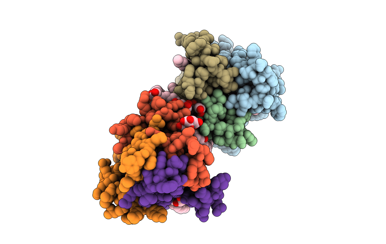
4UYO image
Entry Detail
PDB ID:
4UYO
Keywords:
Title:
Structure of delta7-DgkA in 7.9 MAG by serial femtosecond crystatallography to 2.18 angstrom resolution
Biological Source:
Source Organism:
Host Organism:
PDB Version:
Deposition Date:
2014-09-02
Release Date:
2015-09-30
Method Details:
Experimental Method:
Resolution:
2.18 Å
R-Value Free:
0.23
R-Value Work:
0.20
R-Value Observed:
0.20
Space Group:
P 21 21 21
Macromolecular Entities
Polymer Type:polypeptide(L)
Description:DIACYLGLYCEROL KINASE-DELTA 7
Mutations:YES
Chain IDs:A, B, C, D, E, F
Chain Length:130
Number of Molecules:6
Biological Source:ESCHERICHIA COLI K12
Primary Citation

Abstact

Diacylglycerol kinase catalyses the ATP-dependent conversion of diacylglycerol to phosphatidic acid in the plasma membrane of Escherichia coli. The small size of this integral membrane trimer, which has 121 residues per subunit, means that available protein must be used economically to craft three catalytic and substrate-binding sites centred about the membrane/cytosol interface. How nature has accomplished this extraordinary feat is revealed here in a crystal structure of the kinase captured as a ternary complex with bound lipid substrate and an ATP analogue. Residues, identified as essential for activity by mutagenesis, decorate the active site and are rationalized by the ternary structure. The γ-phosphate of the ATP analogue is positioned for direct transfer to the primary hydroxyl of the lipid whose acyl chain is in the membrane. A catalytic mechanism for this unique enzyme is proposed. The active site architecture shows clear evidence of having arisen by convergent evolution.

Legend

Protein

Chemical

Disease

Primary Citation of related structures