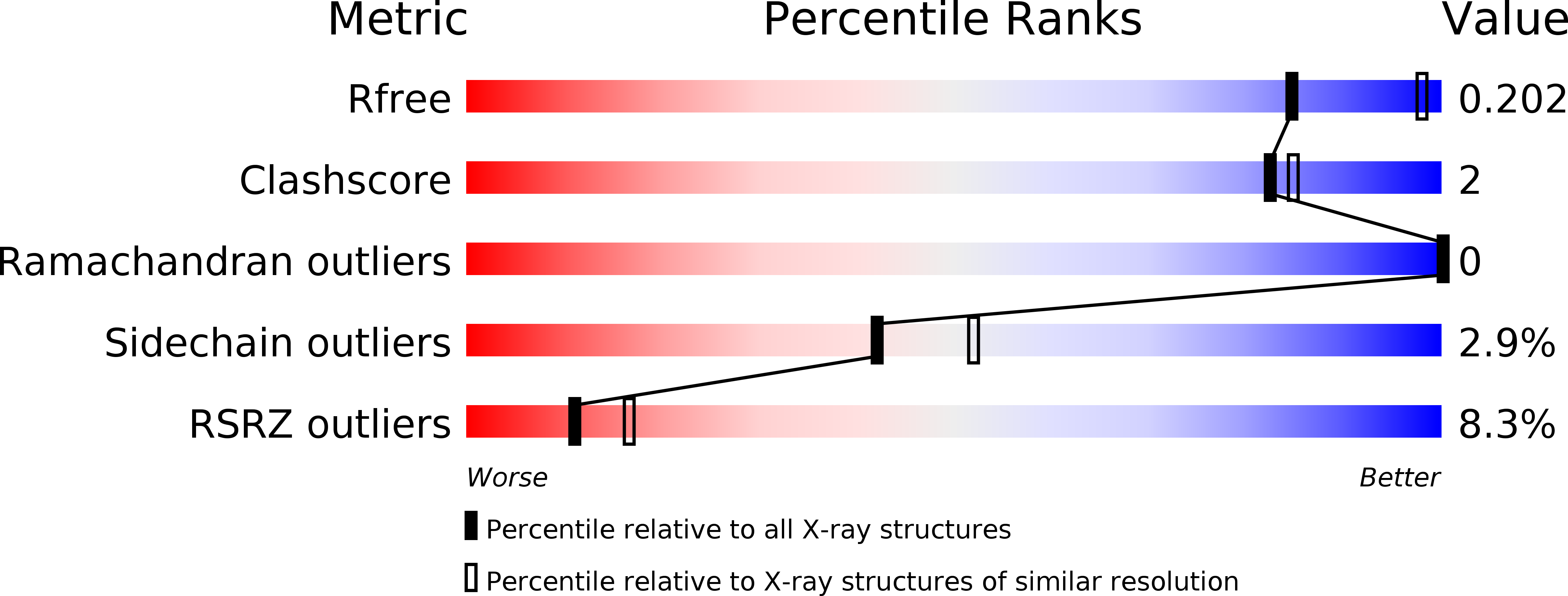
4UX9 image
Deposition Date 2014-08-20
Release Date 2015-03-25
Last Version Date 2024-01-10
Entry Detail
PDB ID:
4UX9
Keywords:
Title:
Crystal structure of JNK1 bound to a MKK7 docking motif
Biological Source:
Source Organism:
HOMO SAPIENS (Taxon ID: 9606)
Host Organism:
Method Details:
Experimental Method:
Resolution:
2.34 Å
R-Value Free:
0.24
R-Value Work:
0.19
R-Value Observed:
0.20
Space Group:
C 1 2 1
Macromolecular Entities
Structures with similar UniProt ID
Protein Blast
Polymer Type:polypeptide(L)
Molecule:MITOGEN-ACTIVATED PROTEIN KINASE 8
Gene (Uniprot):MAPK8
Chain IDs:A, B, C, D
Chain Length:364
Number of Molecules:4
Biological Source:HOMO SAPIENS
Structures with similar UniProt ID
Protein Blast
Polymer Type:polypeptide(L)
Molecule:DUAL SPECIFICITY MITOGEN-ACTIVATED PROTEIN KINASE KINASE 7
Gene (Uniprot):MAP2K7
Chain IDs:E (auth: F), F (auth: G), G (auth: H), H (auth: I)
Chain Length:12
Number of Molecules:4
Biological Source:HOMO SAPIENS
Primary Citation
Structure and Dynamics of the Mkk7-Jnk Signaling Complex.
Proc.Natl.Acad.Sci.USA 112 3409 ? (2015)
PMID: 25737554 DOI: 10.1073/PNAS.1419528112

Abstact

Signaling specificity in the mitogen-activated protein kinase (MAPK) pathways is controlled by disordered domains of the MAPK kinases (MKKs) that specifically bind to their cognate MAPKs via linear docking motifs. MKK7 activates the c-Jun N-terminal kinase (JNK) pathway and is the only MKK containing three motifs within its regulatory domain. Here, we characterize the conformational behavior and interaction mechanism of the MKK7 regulatory domain. Using NMR spectroscopy, we develop an atomic resolution ensemble description of MKK7, revealing highly diverse intrinsic conformational propensities of the three docking sites, suggesting that prerecognition sampling of the bound-state conformation is not prerequisite for binding. Although the different sites exhibit similar affinities for JNK1, interaction kinetics differ considerably. Importantly, we determine the crystal structure of JNK1 in complex with the second docking site of MKK7, revealing two different binding modes of the docking motif correlating with observations from NMR exchange spectroscopy. Our results provide unique insight into how signaling specificity is regulated by linear motifs and, in general, into the role of conformational disorder in MAPK signaling.

Legend

Protein

Chemical

Disease

Primary Citation of related structures