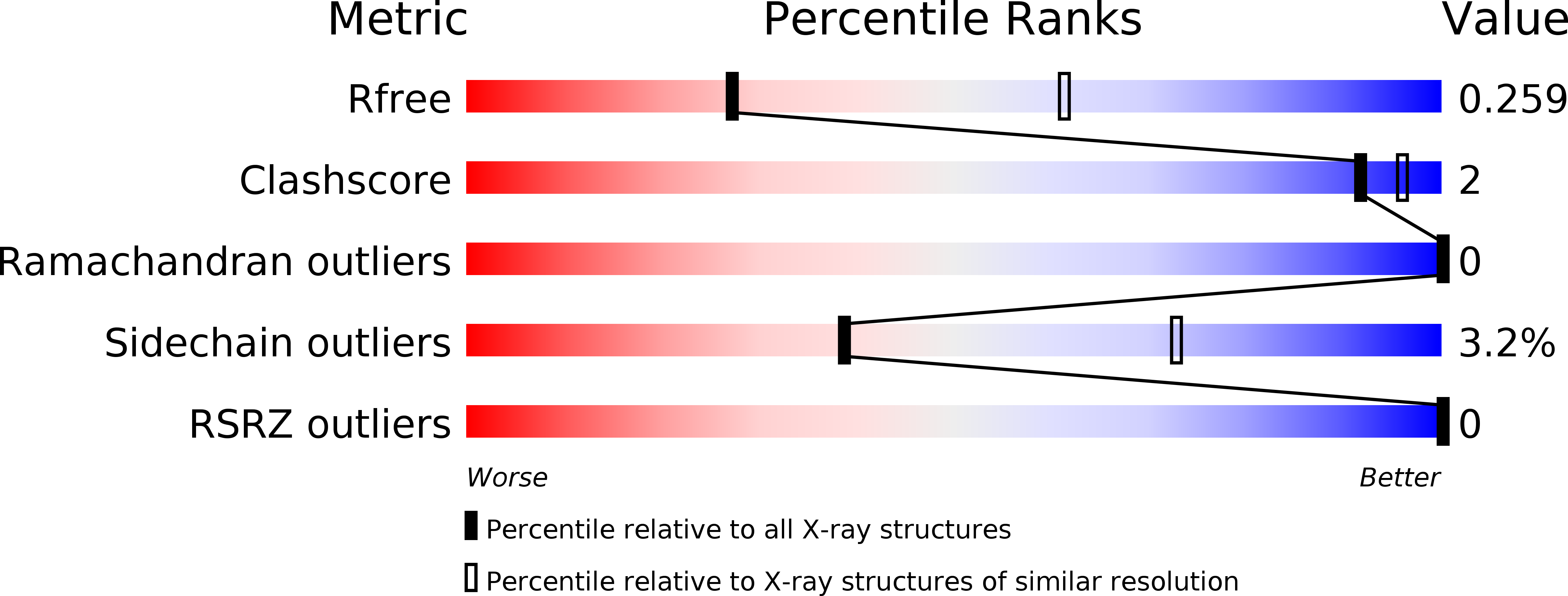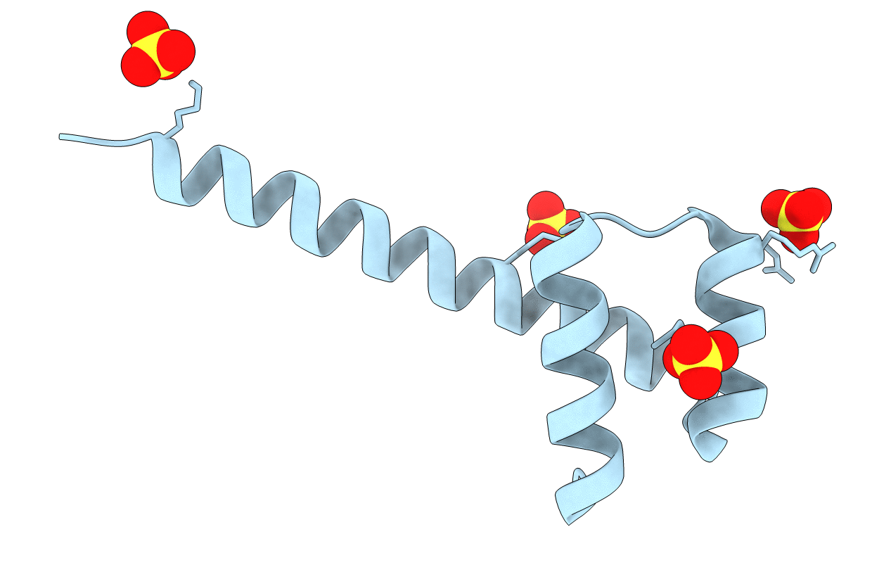
4UUT image
Deposition Date 2014-07-31
Release Date 2015-02-18
Last Version Date 2024-05-01
Entry Detail
PDB ID:
4UUT
Keywords:
Title:
Crystal structure of the Ultrabithorax protein
Biological Source:
Source Organism:
Host Organism:
Method Details:
Experimental Method:
Resolution:
2.80 Å
R-Value Free:
0.23
R-Value Work:
0.21
R-Value Observed:
0.21
Space Group:
P 41 3 2
Macromolecular Entities
Structures with similar UniProt ID
Protein Blast
Polymer Type:polypeptide(L)
Molecule:HOMEOTIC PROTEIN ULTRABITHORAX
Gene (Uniprot):Ubx
Chain IDs:A
Chain Length:93
Number of Molecules:1
Biological Source:DROSOPHILA MELANOGASTER
Primary Citation
A Flexible Extension of the Drosophila Ultrabithorax Homeodomain Defines a Novel Hox/Pbc Interaction Mode.
Structure 23 270 ? (2015)
PMID: 25651060 DOI: 10.1016/J.STR.2014.12.011

Abstact

The patterning function of Hox proteins relies on assembling protein complexes with PBC proteins, which often involves a protein motif found in most Hox proteins, the so-called Hexapeptide (HX). Hox/PBC complexes likely gained functional diversity by acquiring additional modes of interaction. Here, we structurally characterize the first HX alternative interaction mode based on the paralogue-specific UbdA motif and further functionally validate structure-based predictions. The UbdA motif folds as a flexible extension of the homeodomain recognition helix and defines Hox/PBC contacts that occur, compared with those mediated by the HX motif, on the opposing side of the DNA double helix. This provides a new molecular facet to Hox/PBC complex assembly and suggests possible mechanisms for the diversification of Hox protein function.

Legend

Protein

Chemical

Disease

Primary Citation of related structures