4UU5 image
Deposition Date 2014-07-24
Release Date 2015-01-14
Last Version Date 2024-01-10
Entry Detail
PDB ID:
4UU5
Keywords:
Title:
CRYSTAL STRUCTURE OF THE PDZ DOMAIN OF PALS1 IN COMPLEX WITH THE CRB PEPTIDE
Biological Source:
Source Organism:
HOMO SAPIENS (Taxon ID: 9606)
Host Organism:
Method Details:
Experimental Method:
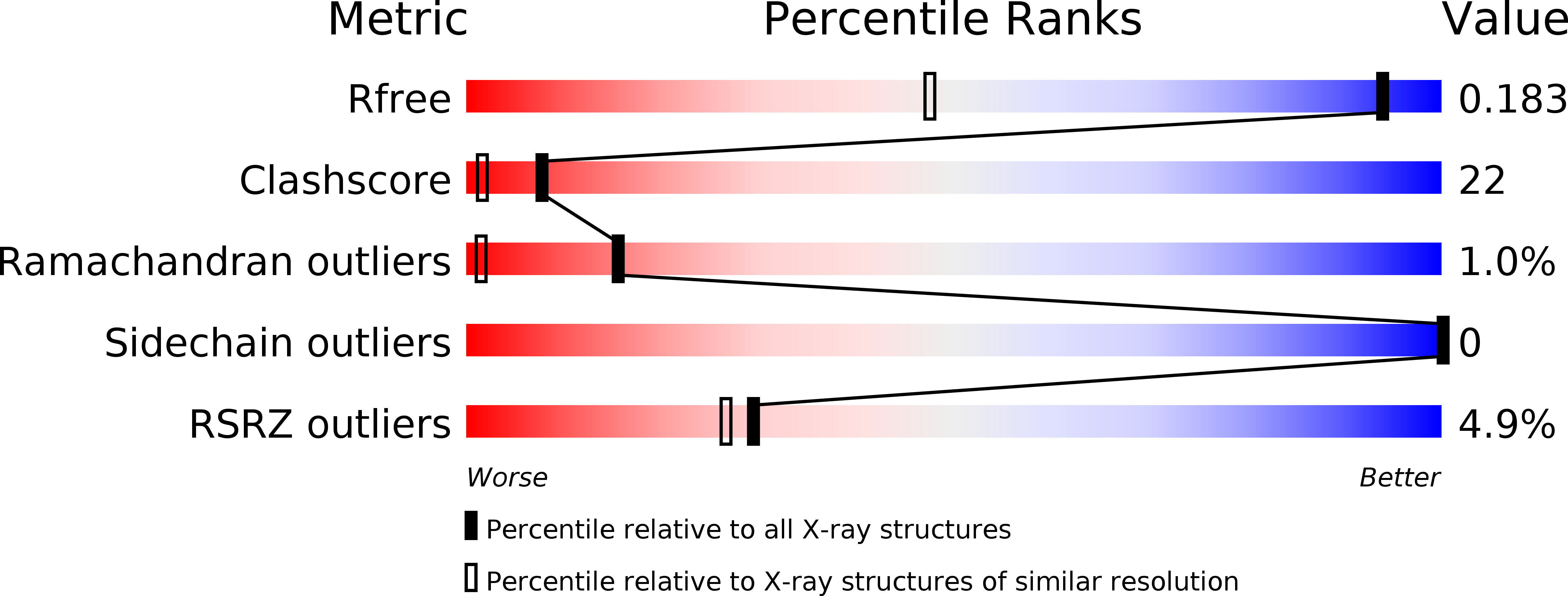
Resolution:
1.23 Å
R-Value Free:
0.18
R-Value Work:
0.16
R-Value Observed:
0.16
Space Group:
P 41 2 2
Macromolecular Entities
Structures with similar UniProt ID
Protein Blast
Polymer Type:polypeptide(L)
Molecule:MAGUK P55 SUBFAMILY MEMBER 5
Gene (Uniprot):PALS1
Chain IDs:A
Chain Length:90
Number of Molecules:1
Biological Source:HOMO SAPIENS
Structures with similar UniProt ID
Protein Blast
Polymer Type:polypeptide(L)
Molecule:PROTEIN CRUMBS HOMOLOG 1
Gene (Uniprot):CRB1
Chain IDs:B
Chain Length:17
Number of Molecules:1
Biological Source:HOMO SAPIENS
Modified Residue
Compound ID Chain ID Parent Comp ID Details 2D Image
SME B MET METHIONINE SULFOXIDE
Primary Citation
Structures of the human Pals1 PDZ domain with and without ligand suggest gated access of Crb to the PDZ peptide-binding groove.
Acta Crystallogr. D Biol. Crystallogr. 71 555 564 (2015)
PMID: 25760605 DOI: 10.1107/S139900471402776X

Abstact

Many components of epithelial polarity protein complexes possess PDZ domains that are required for protein interaction and recruitment to the apical plasma membrane. Apical localization of the Crumbs (Crb) transmembrane protein requires a PDZ-mediated interaction with Pals1 (protein-associated with Lin7, Stardust, MPP5), a member of the p55 family of membrane-associated guanylate kinases (MAGUKs). This study describes the molecular interaction between the Crb carboxy-terminal motif (ERLI), which is required for Drosophila cell polarity, and the Pals1 PDZ domain using crystallography and fluorescence polarization. Only the last four Crb residues contribute to Pals1 PDZ-domain binding affinity, with specificity contributed by conserved charged interactions. Comparison of the Crb-bound Pals1 PDZ structure with an apo Pals1 structure reveals a key Phe side chain that gates access to the PDZ peptide-binding groove. Removal of this side chain enhances the binding affinity by more than fivefold, suggesting that access of Crb to Pals1 may be regulated by intradomain contacts or by protein-protein interaction.

Legend

Protein

Chemical

Disease

Primary Citation of related structures
Feedback Form
Name
Email
Institute
Feedback