4UT0 image
Deposition Date 2014-07-17
Release Date 2014-12-17
Last Version Date 2024-01-10
Entry Detail
PDB ID:
4UT0
Keywords:
Title:
THE CRYSTAL STRUCTURE OF I-DMOI IN COMPLEX WITH ITS TARGET DNA AT 10 DAYS INCUBATION IN 5MM MN (STATE 7)
Biological Source:
Source Organism:
Host Organism:
Method Details:
Experimental Method:
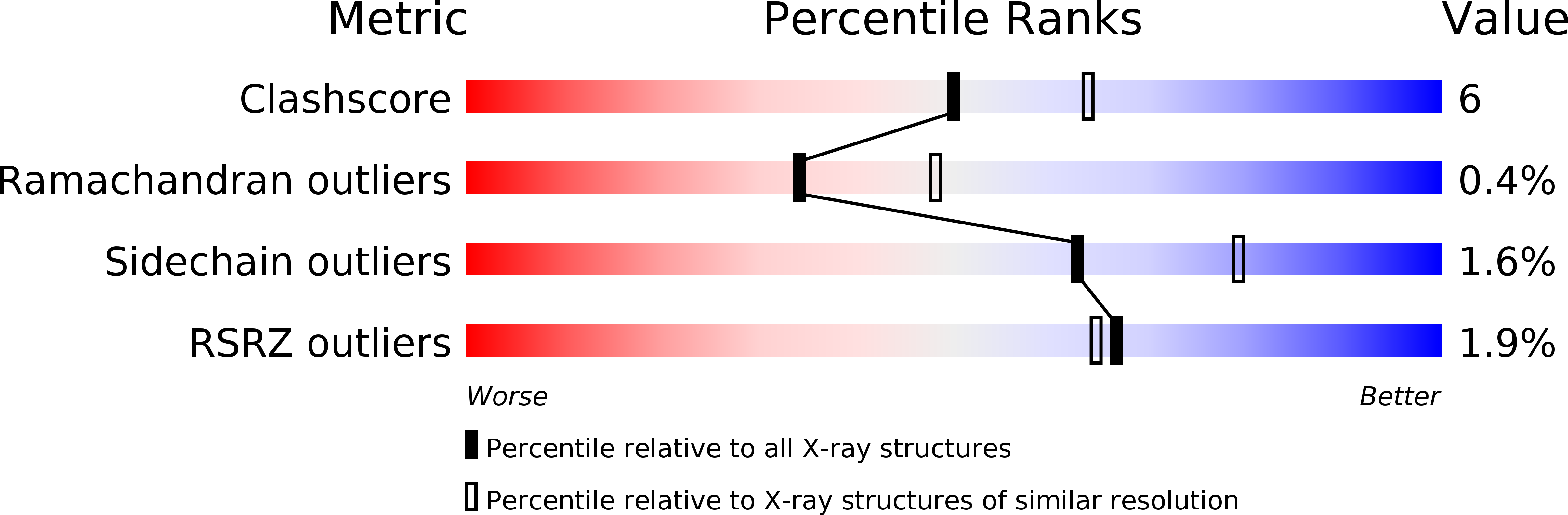
Resolution:
2.40 Å
R-Value Free:
0.22
R-Value Work:
0.18
R-Value Observed:
0.18
Space Group:
P 1 21 1
Macromolecular Entities
Polymer Type:polypeptide(L)
Molecule:HOMING ENDONUCLEASE I-DMOI
Chain IDs:A, F, K
Chain Length:199
Number of Molecules:3
Biological Source:DESULFUROCOCCUS MOBILIS
Polymer Type:polydeoxyribonucleotide
Molecule:5'-D(*GP*CP*CP*TP*TP*GP*CP*CP*GP*GP*GP*TP*AP*AP)-3'
Chain IDs:B, G, L
Chain Length:14
Number of Molecules:3
Biological Source:SYNTHETIC CONSTRUCT
Polymer Type:polydeoxyribonucleotide
Molecule:5'-D(*GP*TP*TP*CP*CP*GP*GP*CP*GP*CP*GP)-3'
Chain IDs:C, H, M
Chain Length:11
Number of Molecules:3
Biological Source:SYNTHETIC CONSTRUCT
Polymer Type:polydeoxyribonucleotide
Molecule:5'-D(*CP*GP*CP*GP*CP*CP*GP*GP*AP*AP*CP*TP*TP*AP*CP)-3'
Chain IDs:D, I, N
Chain Length:15
Number of Molecules:3
Biological Source:SYNTHETIC CONSTRUCT
Polymer Type:polydeoxyribonucleotide
Molecule:5'-D(*CP*CP*GP*GP*CP*AP*AP*GP*GP*CP)-3'
Chain IDs:E, J, O
Chain Length:10
Number of Molecules:3
Biological Source:SYNTHETIC CONSTRUCT
Primary Citation
Visualizing Phosphodiester-Bond Hydrolysis by an Endonuclease.
Nat.Struct.Mol.Biol. 22 65 ? (2015)
PMID: 25486305 DOI: 10.1038/NSMB.2932

Abstact

The enzymatic hydrolysis of DNA phosphodiester bonds has been widely studied, but the chemical reaction has not yet been observed. Here we follow the generation of a DNA double-strand break (DSB) by the Desulfurococcus mobilis homing endonuclease I-DmoI, trapping sequential stages of a two-metal-ion cleavage mechanism. We captured intermediates of the different catalytic steps, and this allowed us to watch the reaction by 'freezing' multiple states. We observed the successive entry of two metals involved in the reaction and the arrival of a third cation in a central position of the active site. This third metal ion has a crucial role, triggering the consecutive hydrolysis of the targeted phosphodiester bonds in the DNA strands and leaving its position once the DSB is generated. The multiple structures show the orchestrated conformational changes in the protein residues, nucleotides and metals during catalysis.

Legend

Protein

Chemical

Disease

Primary Citation of related structures