Deposition Date
2014-06-30
Release Date
2014-07-30
Last Version Date
2024-05-08
Entry Detail
PDB ID:
4URM
Keywords:
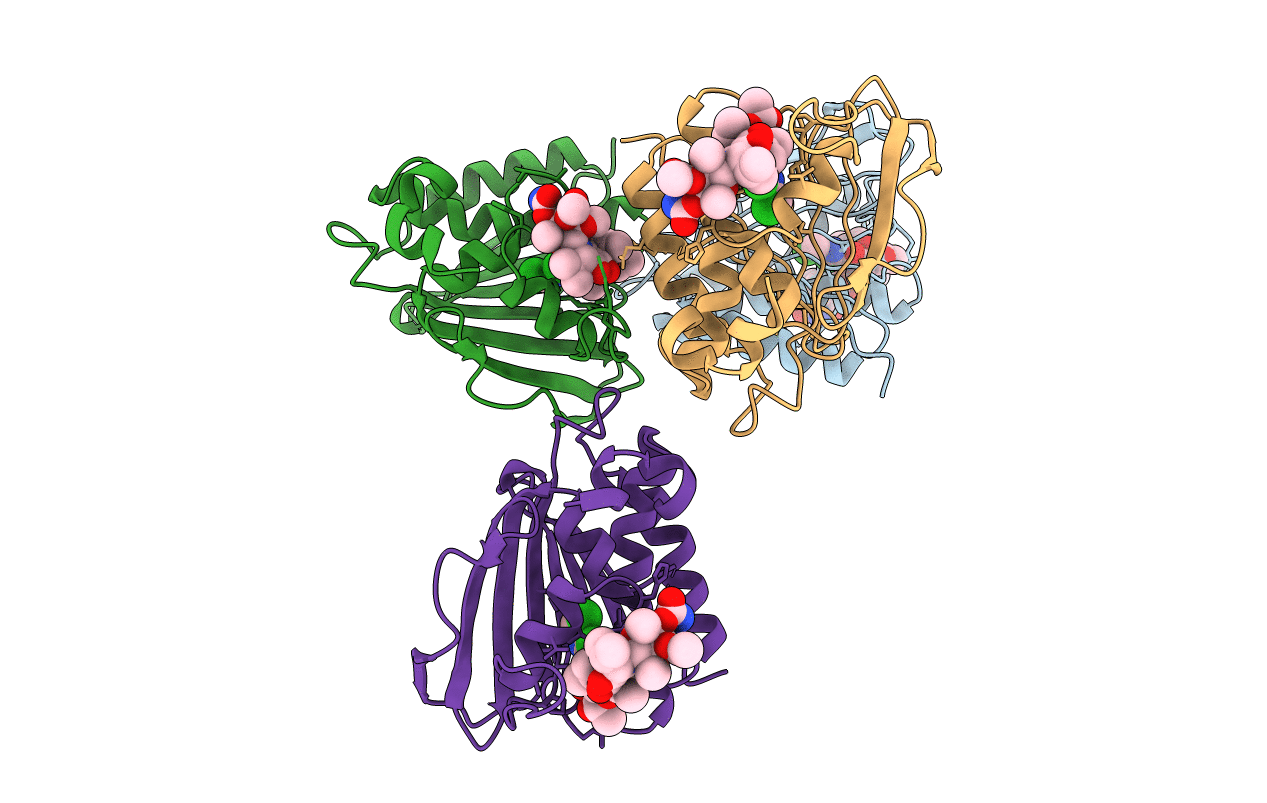
Title:
Crystal Structure of Staph GyraseB 24kDa in complex with Kibdelomycin
Biological Source:
Source Organism(s):
STAPHYLOCOCCUS AUREUS (Taxon ID: 1280)
Expression System(s):
Method Details:
Experimental Method:
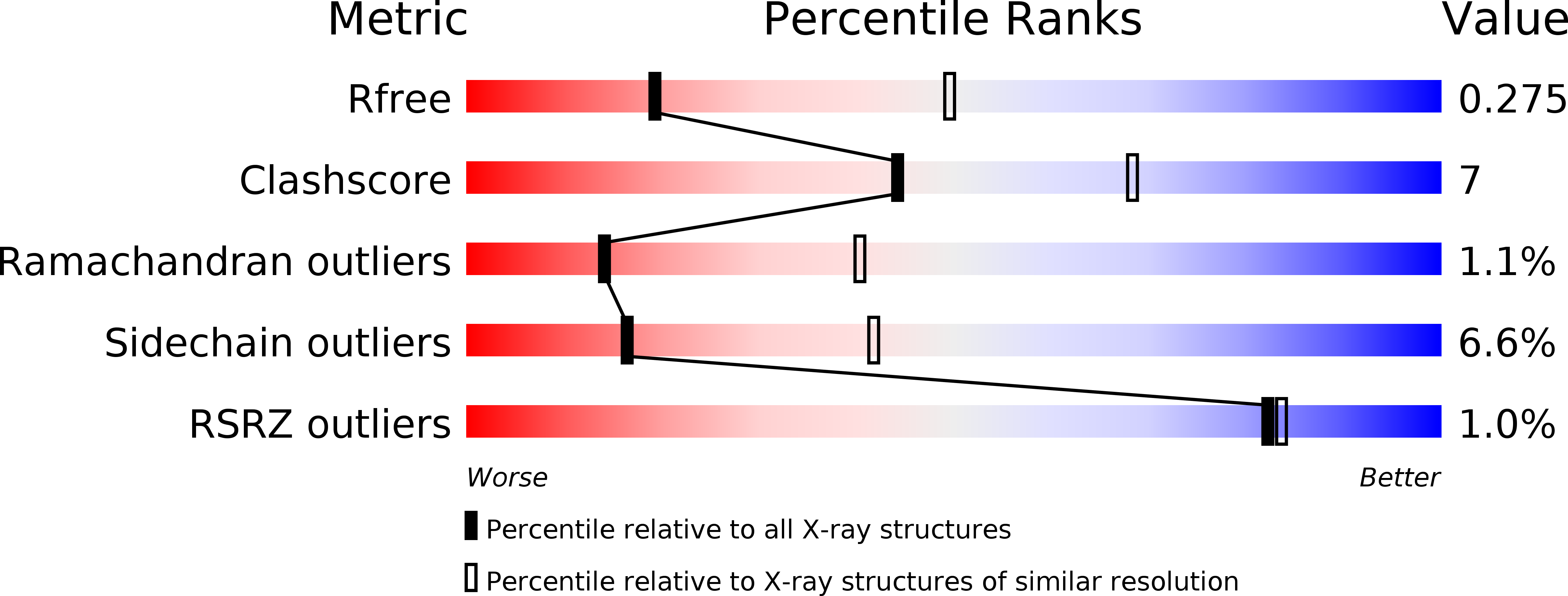
Resolution:
2.94 Å
R-Value Free:
0.25
R-Value Work:
0.19
R-Value Observed:
0.19
Space Group:
P 1 21 1