4UQM image
Deposition Date 2014-06-24
Release Date 2015-08-12
Last Version Date 2024-01-10
Entry Detail
PDB ID:
4UQM
Keywords:
Title:
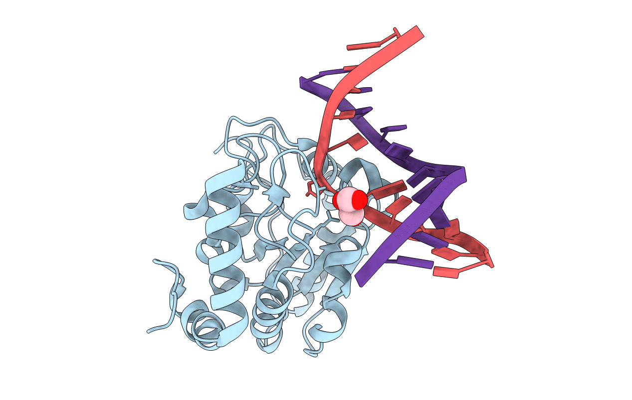
Crystal structure determination of uracil-DNA N-glycosylase (UNG) from Deinococcus radiodurans in complex with DNA - new insights into the role of the Leucine-loop for damage recognition and repair
Biological Source:
Source Organism:
Host Organism:
Method Details:
Experimental Method:
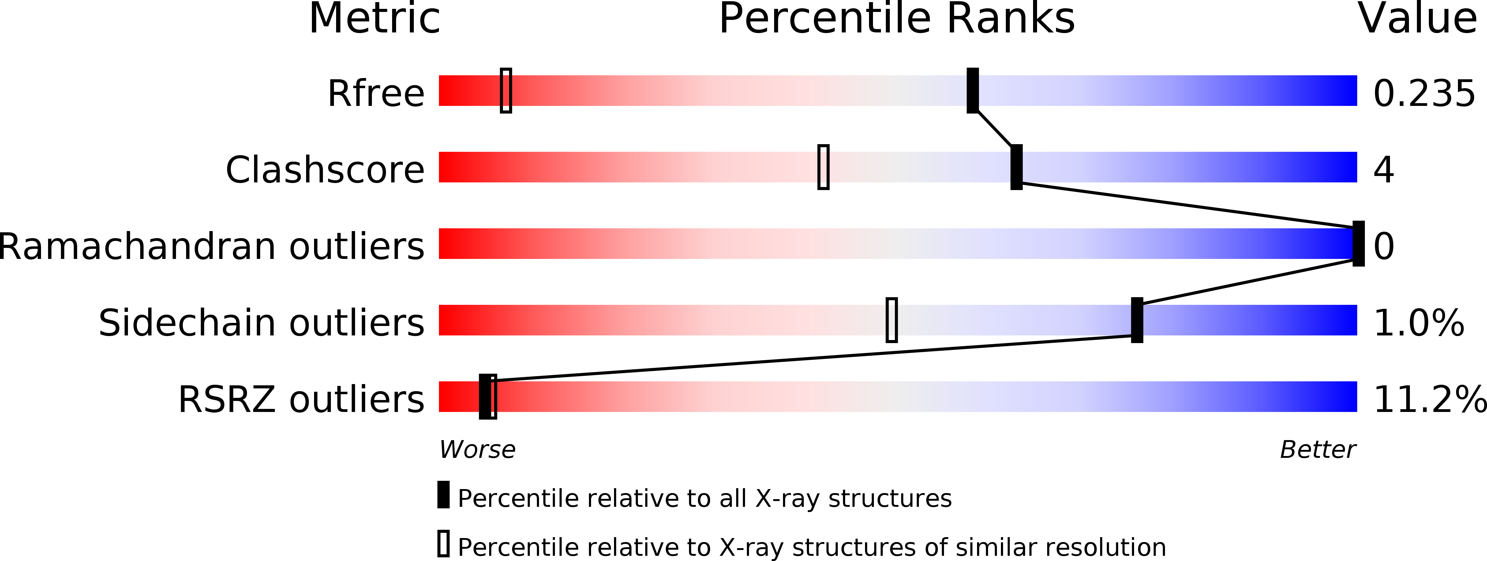
Resolution:
1.35 Å
R-Value Free:
0.21
R-Value Work:
0.18
R-Value Observed:
0.18
Space Group:
P 21 21 2
Macromolecular Entities
Structures with similar UniProt ID
Protein Blast
Polymer Type:polypeptide(L)
Molecule:URACIL-DNA GLYCOSYLASE
Gene (Uniprot):ung
Mutagens:YES
Chain IDs:A
Chain Length:247
Number of Molecules:1
Biological Source:DEINOCOCCUS RADIODURANS
Polymer Type:polydeoxyribonucleotide
Molecule:5'-D(*CP*CP*TP*AP*TP*CP*CP*AP*AAB*GP*TP*CP*TP*CP*CP*G)-3'
Chain IDs:B
Chain Length:16
Number of Molecules:1
Biological Source:SYNTHETIC CONSTRUCT
Polymer Type:polydeoxyribonucleotide
Molecule:5'-D(*GP*CP*GP*GP*AP*GP*AP*CP*AP*TP*GP*GP*AP*CP*AP*G)-3'
Chain IDs:C
Chain Length:16
Number of Molecules:1
Biological Source:SYNTHETIC CONSTRUCT
Primary Citation
Structure determination of uracil-DNA N-glycosylase from Deinococcus radiodurans in complex with DNA.
Acta Crystallogr. D Biol. Crystallogr. 71 2137 2149 (2015)
PMID: 26457437 DOI: 10.1107/S1399004715014157

Abstact

Uracil-DNA N-glycosylase (UNG) is a DNA-repair enzyme in the base-excision repair (BER) pathway which removes uracil from DNA. Here, the crystal structure of UNG from the extremophilic bacterium Deinococcus radiodurans (DrUNG) in complex with DNA is reported at a resolution of 1.35 Å. Prior to the crystallization experiments, the affinity between DrUNG and different DNA oligonucleotides was tested by electrophoretic mobility shift assays (EMSAs). As a result of this analysis, two 16 nt double-stranded DNAs were chosen for the co-crystallization experiments, one of which (16 nt AU) resulted in well diffracting crystals. The DNA in the co-crystal structure contained an abasic site (substrate product) flipped into the active site of the enzyme, with no uracil in the active-site pocket. Despite the high resolution, it was not possible to fit all of the terminal nucleotides of the DNA complex into electron density owing to disorder caused by a lack of stabilizing interactions. However, the DNA which was in contact with the enzyme, close to the active site, was well ordered and allowed detailed analysis of the enzyme-DNA interaction. The complex revealed that the interaction between DrUNG and DNA is similar to that in the previously determined crystal structure of human UNG (hUNG) in complex with DNA [Slupphaug et al. (1996). Nature (London), 384, 87-92]. Substitutions in a (here defined) variable part of the leucine loop result in a shorter loop (eight residues instead of nine) in DrUNG compared with hUNG; regardless of this, it seems to fulfil its role and generate a stabilizing force with the minor groove upon flipping out of the damaged base into the active site. The structure also provides a rationale for the previously observed high catalytic efficiency of DrUNG caused by high substrate affinity by demonstrating an increased number of long-range electrostatic interactions between the enzyme and the DNA. Interestingly, specific interactions between residues in the N-terminus of a symmetry-related molecule and the complementary DNA strand facing away from the active site were also observed which seem to stabilize the enzyme-DNA complex. However, the significance of this observation remains to be investigated. The results provide new insights into the current knowledge about DNA damage recognition and repair by uracil-DNA glycosylases.

Legend

Protein

Chemical

Disease

Primary Citation of related structures