4UM7 image
Entry Detail
PDB ID:
4UM7
Keywords:
Title:
Crystal structure of 3-deoxy-D-manno-octulosonate 8-phosphate phosphatase (kdsC) from Moraxella catarrhalis in complex with Magnesium ion
Biological Source:
Source Organism:
Host Organism:
PDB Version:
Deposition Date:
2014-05-15
Release Date:
2015-02-11
Method Details:
Experimental Method:
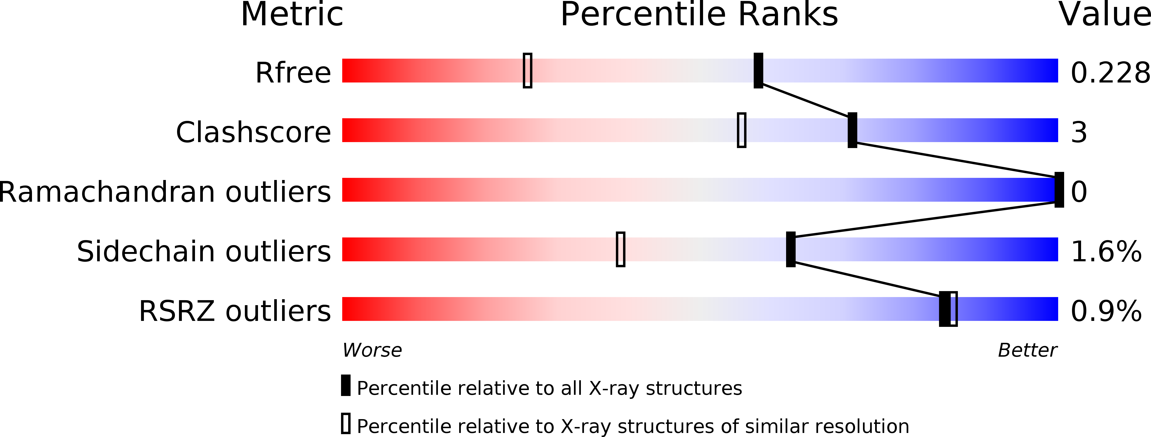
Resolution:
1.64 Å
R-Value Free:
0.22
R-Value Work:
0.19
R-Value Observed:
0.19
Space Group:
P 1 21 1
Macromolecular Entities
Polymer Type:polypeptide(L)
Description:3-DEOXY-D-MANNO-OCTULOSONATE 8-PHOSPHATE PHOSPHATASE KDSC
Chain IDs:A, B, C, D
Chain Length:193
Number of Molecules:4
Biological Source:MORAXELLA CATARRHALIS BC8
Primary Citation
Ligand-Bound Structures of 3-Deoxy-D-Manno-Octulosonate 8-Phosphate Phosphatase from Moraxella Catarrhalis Reveal a Water Channel Connecting to the Active Site for the Second Step of Catalysis
Acta Crystallogr.,Sect.D 71 239 ? (2015)
PMID: 25664734 DOI: 10.1107/S1399004714025218

Abstact

KdsC, the third enzyme of the 3-deoxy-D-manno-octulosonic acid (KDO) biosynthetic pathway, catalyzes a substrate-specific reaction to hydrolyze 3-deoxy-D-manno-octulosonate 8-phosphate to generate a molecule of KDO and phosphate. KdsC is a phosphatase that belongs to the C0 subfamily of the HAD superfamily. To understand the molecular basis for the substrate specificity of this tetrameric enzyme, the crystal structures of KdsC from Moraxella catarrhalis (Mc-KdsC) with several combinations of ligands, namely metal ion, citrate and products, were determined. Various transition states of the enzyme have been captured in these crystal forms. The ligand-free and ligand-bound crystal forms reveal that the binding of ligands does not cause any specific conformational changes in the active site. However, the electron-density maps clearly showed that the conformation of KDO as a substrate is different from the conformation adopted by KDO when it binds as a cleaved product. Furthermore, structural evidence for the existence of an intersubunit tunnel has been reported for the first time in the C0 subfamily of enzymes. A role for this tunnel in transferring water molecules from the interior of the tetrameric structure to the active-site cleft has been proposed. At the active site, water molecules are required for the formation of a water bridge that participates as a proton shuttle during the second step of the two-step phosphoryl-transfer reaction. In addition, as the KDO biosynthesis pathway is a potential antibacterial target, pharmacophore-based virtual screening was employed to identify inhibitor molecules for the Mc-KdsC enzyme.

Legend

Protein

Chemical

Disease

Primary Citation of related structures