Deposition Date
2014-12-10
Release Date
2015-05-27
Last Version Date
2023-12-20
Entry Detail
PDB ID:
4UDJ
Keywords:
Title:
Crystal structure of b-1,4-mannopyranosyl-chitobiose phosphorylase at 1.60 Angstrom in complex with beta-D-mannopyranose and inorganic phosphate
Biological Source:
Source Organism(s):
UNCULTURED ORGANISM (Taxon ID: 155900)
Expression System(s):
Method Details:
Experimental Method:
Resolution:
1.94 Å
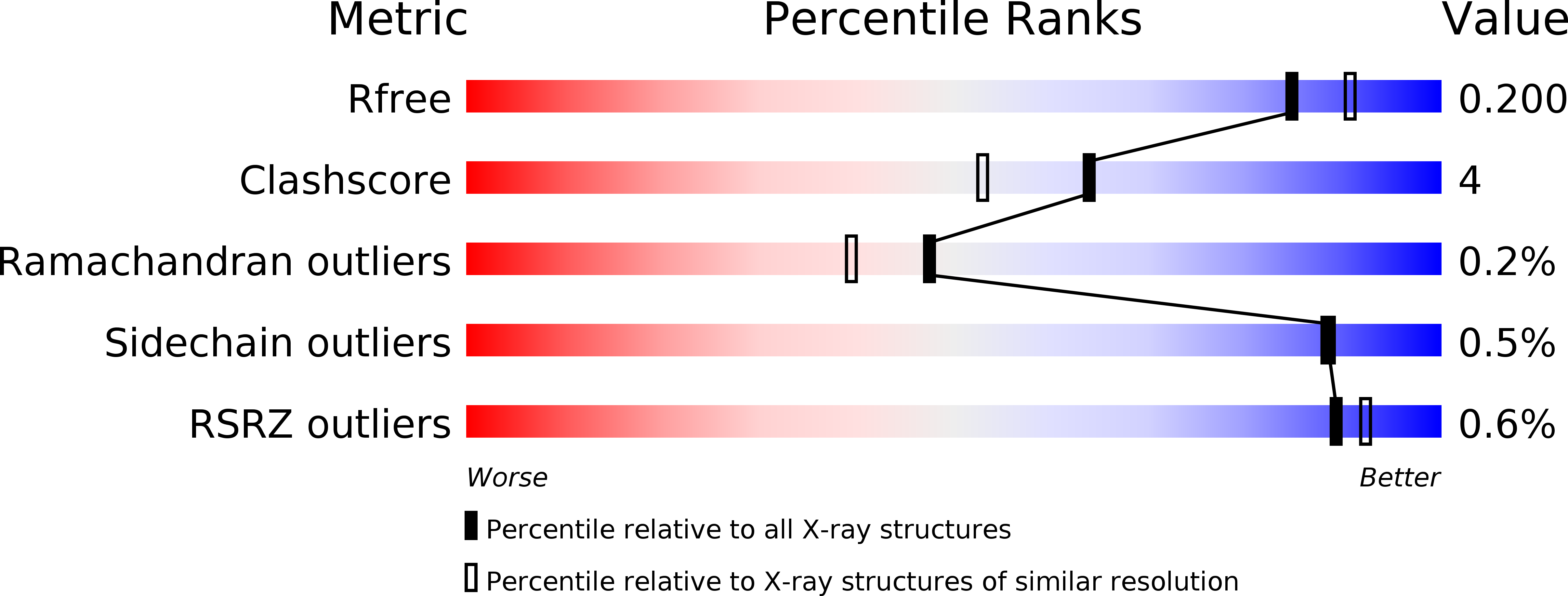
R-Value Free:
0.19
R-Value Work:
0.14
R-Value Observed:
0.15
Space Group:
P 21 21 21