4UA9 image
Deposition Date 2014-08-08
Release Date 2015-06-24
Last Version Date 2024-11-06
Entry Detail
PDB ID:
4UA9
Keywords:
Title:
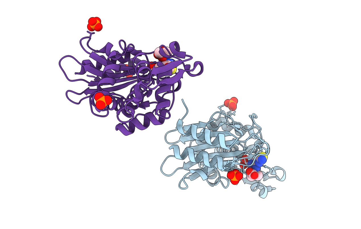
CTX-M-14 Class A Beta-Lactamase in Complex with a Boronic Acid Acylation Transition State Analog at Sub-Angstrom Resolution
Biological Source:
Source Organism:
Escherichia coli (Taxon ID: 562)
Host Organism:
Method Details:
Experimental Method:
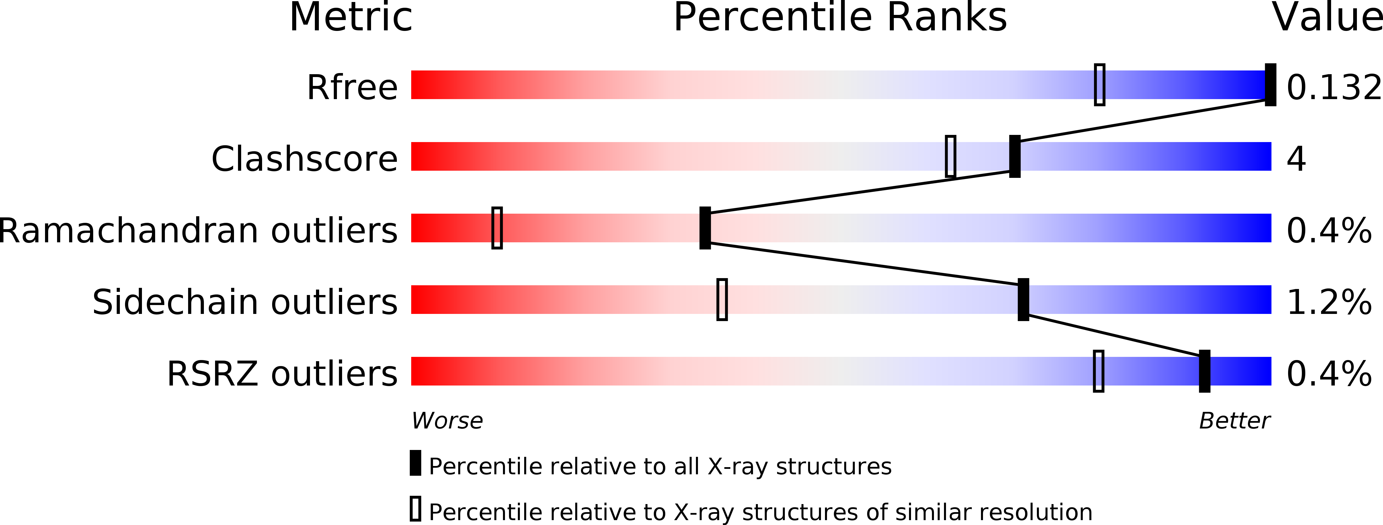
Resolution:
0.84 Å
R-Value Free:
0.13
R-Value Work:
0.12
R-Value Observed:
0.12
Space Group:
P 1 21 1
Macromolecular Entities
Structures with similar UniProt ID
Protein Blast
Polymer Type:polypeptide(L)
Molecule:Beta-lactamase CTX-M-14
Gene (Uniprot):CTX-M-14
Chain IDs:A, B
Chain Length:263
Number of Molecules:2
Biological Source:Escherichia coli
Primary Citation
Ligand-Induced Proton Transfer and Low-Barrier Hydrogen Bond Revealed by X-ray Crystallography.
J.Am.Chem.Soc. 137 8086 8095 (2015)
PMID: 26057252 DOI: 10.1021/jacs.5b00749

Abstact

Ligand binding can change the pKa of protein residues and influence enzyme catalysis. Herein, we report three ultrahigh resolution X-ray crystal structures of CTX-M β-lactamase, directly visualizing protonation state changes along the enzymatic pathway: apo protein at 0.79 Å, precovalent complex with nonelectrophilic ligand at 0.89 Å, and acylation transition state (TS) analogue at 0.84 Å. Binding of the noncovalent ligand induces a proton transfer from the catalytic Ser70 to the negatively charged Glu166, and the formation of a low-barrier hydrogen bond (LBHB) between Ser70 and Lys73, with a length of 2.53 Å and the shared hydrogen equidistant from the heteroatoms. QM/MM reaction path calculations determined the proton transfer barrier to be 1.53 kcal/mol. The LBHB is absent in the other two structures although Glu166 remains neutral in the covalent complex. Our data represents the first X-ray crystallographic example of a hydrogen engaged in an enzymatic LBHB, and demonstrates that desolvation of the active site by ligand binding can provide a protein microenvironment conducive to LBHB formation. It also suggests that LBHBs may contribute to stabilization of the TS in general acid/base catalysis together with other preorganized features of enzyme active sites. These structures reconcile previous experimental results suggesting alternatively Glu166 or Lys73 as the general base for acylation, and underline the importance of considering residue protonation state change when modeling protein-ligand interactions. Additionally, the observation of another LBHB (2.47 Å) between two conserved residues, Asp233 and Asp246, suggests that LBHBs may potentially play a special structural role in proteins.

Legend

Protein

Chemical

Disease

Primary Citation of related structures