4U8T image
Deposition Date 2014-08-04
Release Date 2014-09-10
Last Version Date 2024-10-30
Entry Detail
PDB ID:
4U8T
Title:
Crystal structure of YTH domain of Zygosaccharomyces rouxii MRB1 protein in complex with N6-Methyladenosine RNA
Biological Source:
Source Organism:
Host Organism:
Method Details:
Experimental Method:
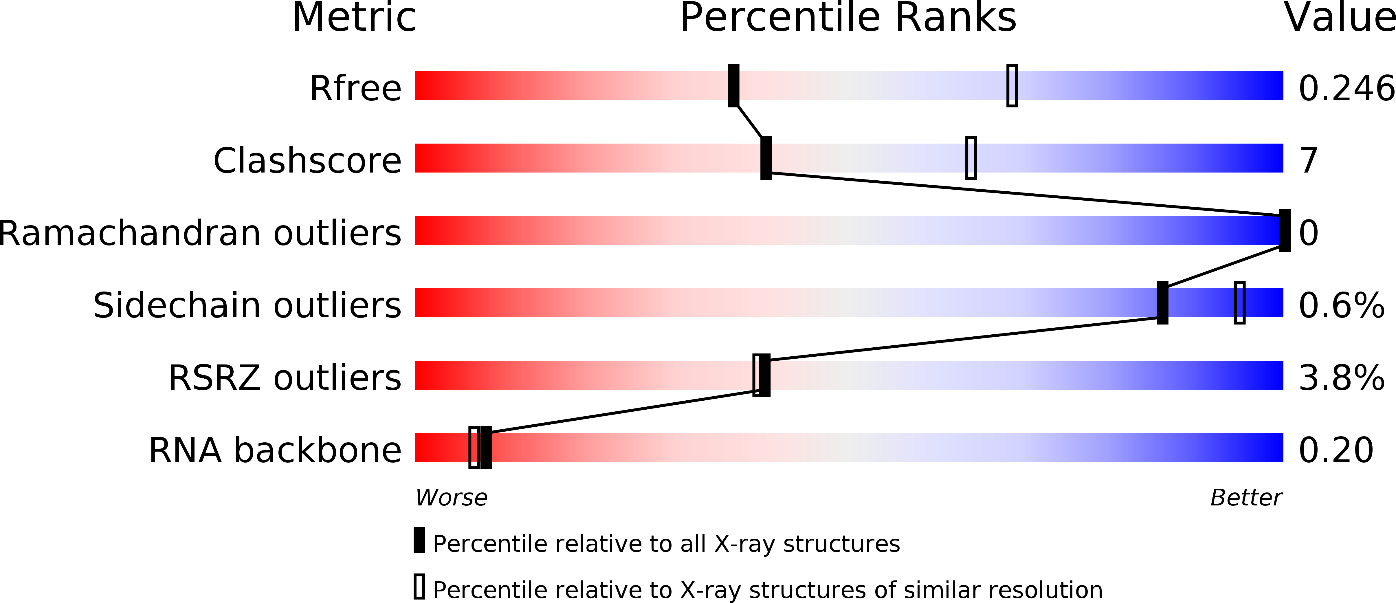
Resolution:
2.70 Å
R-Value Free:
0.24
R-Value Work:
0.19
R-Value Observed:
0.20
Space Group:
P 61 2 2
Macromolecular Entities
Structures with similar UniProt ID
Protein Blast
Polymer Type:polypeptide(L)
Molecule:ZYRO0G01672p
Gene (Uniprot):ZYRO0G01672g
Chain IDs:A, B, C, D, E, F
Chain Length:177
Number of Molecules:6
Biological Source:Zygosaccharomyces rouxii
Polymer Type:polyribonucleotide
Molecule:RNA (5'-R(*AP*GP*GP*(6MZ)P*CP*AP*U)-3')
Chain IDs:G (auth: O), H (auth: P), I (auth: Q), J (auth: R), K (auth: S), L (auth: T)
Chain Length:7
Number of Molecules:6
Biological Source:Saccharomyces cerevisiae
Modified Residue
Compound ID Chain ID Parent Comp ID Details 2D Image
MSE A MET modified residue
Primary Citation
Molecular basis for the recognition of methylated adenines in RNA by the eukaryotic YTH domain.
Proc.Natl.Acad.Sci.USA 111 13834 13839 (2014)
PMID: 25201973 DOI: 10.1073/pnas.1412742111

Abstact

Methylation of the N6 position of selected internal adenines (m(6)A) in mRNAs and noncoding RNAs is widespread in eukaryotes, and the YTH domain in a collection of proteins recognizes this modification. We report the crystal structure of the splicing factor YT521-B homology (YTH) domain of Zygosaccharomyces rouxii MRB1 in complex with a heptaribonucleotide with an m(6)A residue in the center. The m(6)A modification is recognized by an aromatic cage, being sandwiched between a Trp and Tyr residue and with the methyl group pointed toward another Trp residue. Mutations of YTH domain residues in the RNA binding site can abolish the formation of the complex, confirming the structural observations. These residues are conserved in the human YTH proteins that also bind m(6)A RNA, suggesting a conserved mode of recognition. Overall, our structural and biochemical studies have defined the molecular basis for how the YTH domain functions as a reader of methylated adenines.

Legend

Protein

Chemical

Disease

Primary Citation of related structures