4U3T image
Deposition Date 2014-07-22
Release Date 2014-12-03
Last Version Date 2023-09-27
Entry Detail
PDB ID:
4U3T
Keywords:
Title:
Crystal structure of the transpeptidase domain of Neisseria gonorrhoeae penicillin-binding protein 2 derived from the penicillin-resistant strain 6140
Biological Source:
Source Organism:
Host Organism:
Method Details:
Experimental Method:
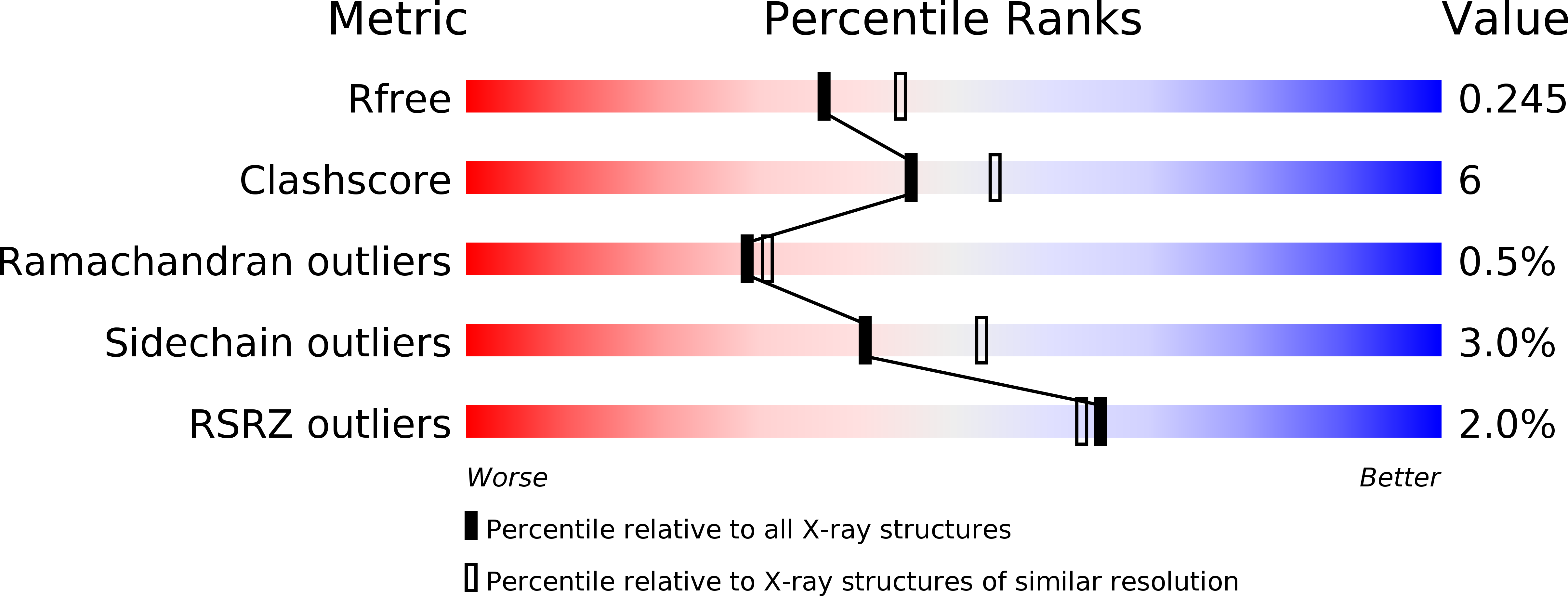
Resolution:
2.20 Å
R-Value Free:
0.24
R-Value Work:
0.19
R-Value Observed:
0.19
Space Group:
P 1 21 1
Macromolecular Entities
Structures with similar UniProt ID
Protein Blast
Polymer Type:polypeptide(L)
Molecule:Penicillin-binding protein 2
Gene (Uniprot):penA
Mutagens:D346a, F504L, A510V, A516G, P551S
Chain IDs:A, B
Chain Length:330
Number of Molecules:2
Biological Source:Neisseria gonorrhoeae FA6140
Primary Citation
Structural Effect of the Asp345a Insertion in Penicillin-Binding Protein 2 from Penicillin-Resistant Strains of Neisseria gonorrhoeae.
Biochemistry 53 7596 7603 (2014)
PMID: 25403720 DOI: 10.1021/bi5011317

Abstact

A hallmark of penicillin-binding protein 2 (PBP2) from penicillin-resistant strains of Neisseria gonorrhoeae is insertion of an aspartate after position 345. The insertion resides on a loop near the active site and is immediately adjacent to an existing aspartate (Asp346) that forms a functionally important hydrogen bond with Ser363 of the SxN conserved motif. Insertion of other amino acids, including Glu and Asn, can also lower the rate of acylation by penicillin, but these insertions abolish transpeptidase function. Although the kinetic consequences of the Asp insertion are well-established, how it impacts the structure of PBP2 is unknown. Here, we report the 2.2 Å resolution crystal structure of a truncated construct of PBP2 containing all five mutations present in PBP2 from the penicillin-resistant strain 6140, including the Asp insertion. Commensurate with the strict specificity for the Asp insertion over similar amino acids, the insertion does not cause disordering of the structure, but rather induces localized flexibility in the β2c-β2d loop. The crystal structure resolves the ambiguity of whether the insertion is Asp345a or Asp346a (due to the adjacent Asp) because the hydrogen bond between Asp346 and Ser362 is preserved and the insertion is therefore Asp346a. The side chain of Asp346a projects directly toward the β-lactam-binding site near Asn364 of the SxN motif. The Asp insertion may lower the rate of acylation by sterically impeding binding of the antibiotic or by hindering breakage of the β-lactam ring during acylation because of the negative charge of its side chain.

Legend

Protein

Chemical

Disease

Primary Citation of related structures
Feedback Form
Name
Email
Institute
Feedback