Deposition Date
2014-07-16
Release Date
2014-12-10
Last Version Date
2023-12-27
Entry Detail
PDB ID:
4U2B
Keywords:
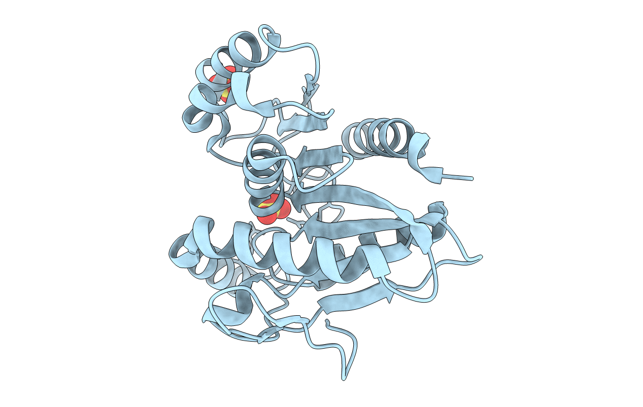
Title:
Crystal structure of dienelactone hydrolase (C123S) at 1.70 A resolution
Biological Source:
Source Organism(s):
Pseudomonas knackmussii (Taxon ID: 65741)
Expression System(s):
Method Details:
Experimental Method:
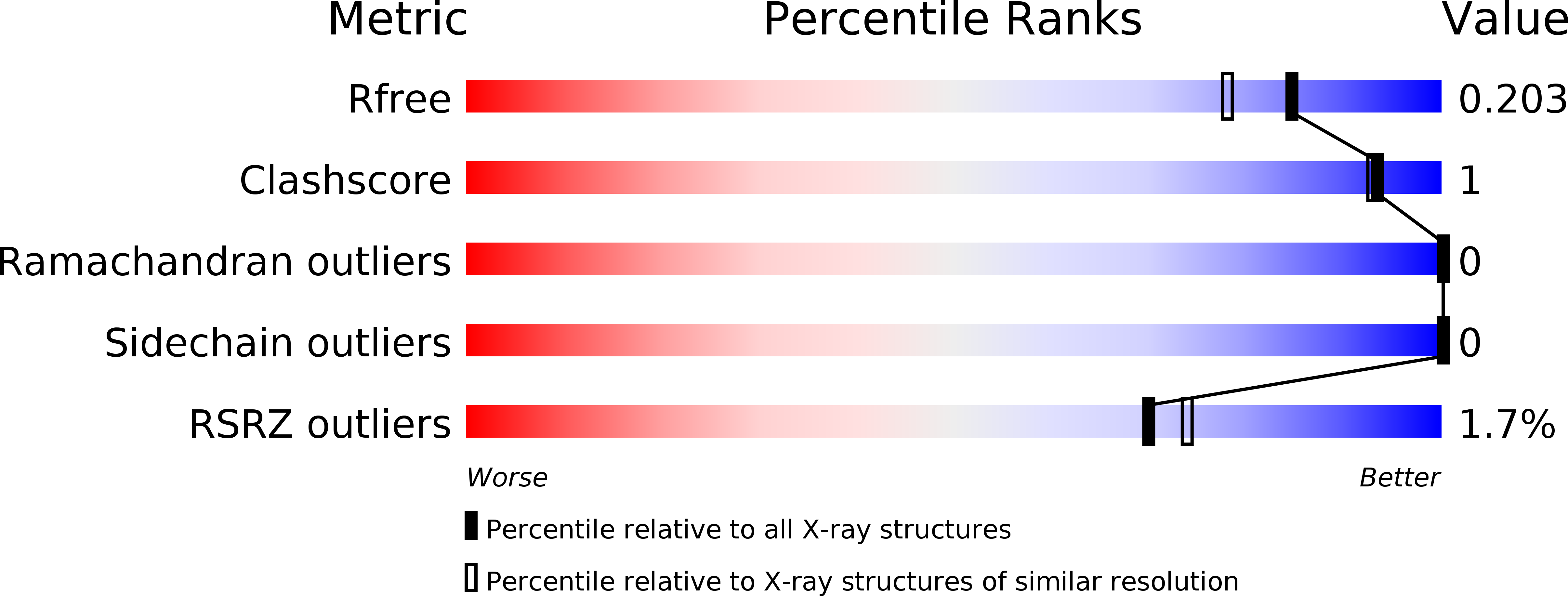
Resolution:
1.70 Å
R-Value Free:
0.19
R-Value Work:
0.17
R-Value Observed:
0.17
Space Group:
P 21 21 21