
Deposition Date
2014-07-15
Release Date
2014-12-24
Last Version Date
2023-12-20
Entry Detail
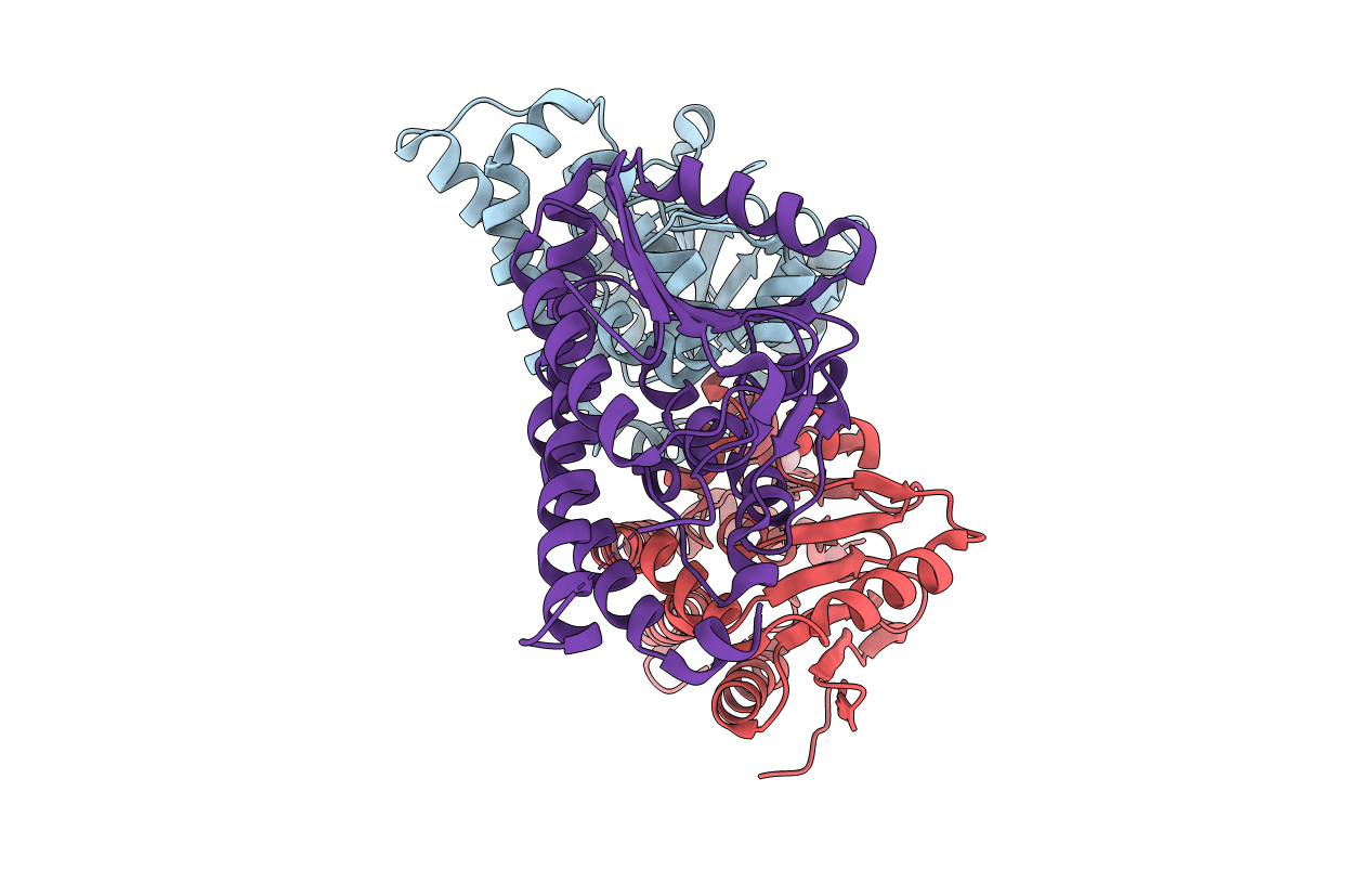
PDB ID:
4U19
Keywords:
Title:
Crystal structure of human peroxisomal delta3,delta2, enoyl-CoA isomerase V349A mutant (ISOA-ECI2)
Biological Source:
Source Organism(s):
Homo sapiens (Taxon ID: 9606)
Expression System(s):
Method Details:
Experimental Method:
Resolution:
1.88 Å
R-Value Free:
0.23
R-Value Work:
0.18
R-Value Observed:
0.18
Space Group:
P 21 21 21
