4U18 image
Deposition Date 2014-07-15
Release Date 2014-12-24
Last Version Date 2024-05-08
Entry Detail
PDB ID:
4U18
Keywords:
Title:
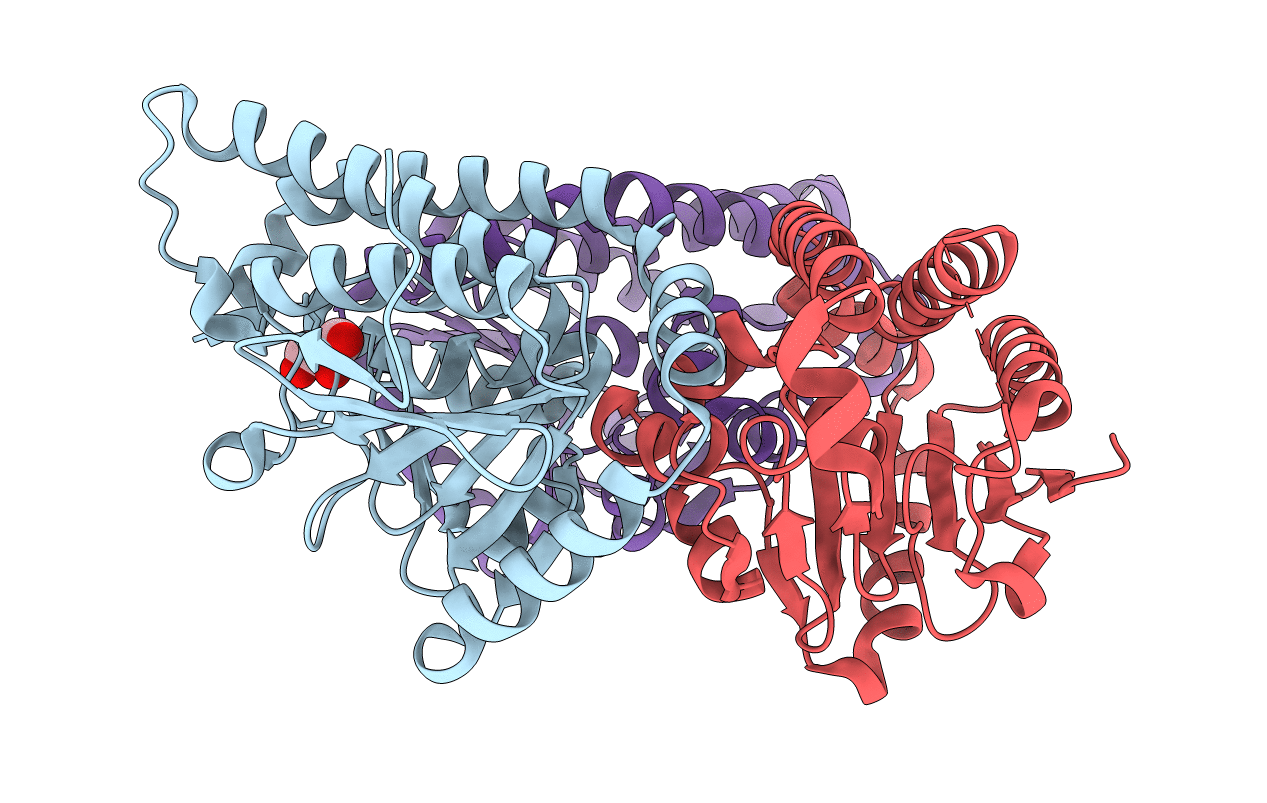
Crystal structure of human peroxisomal delta3,delta2, enoyl-CoA isomerase (ISO-ECI2)
Biological Source:
Source Organism:
Homo sapiens (Taxon ID: 9606)
Host Organism:
Method Details:
Experimental Method:
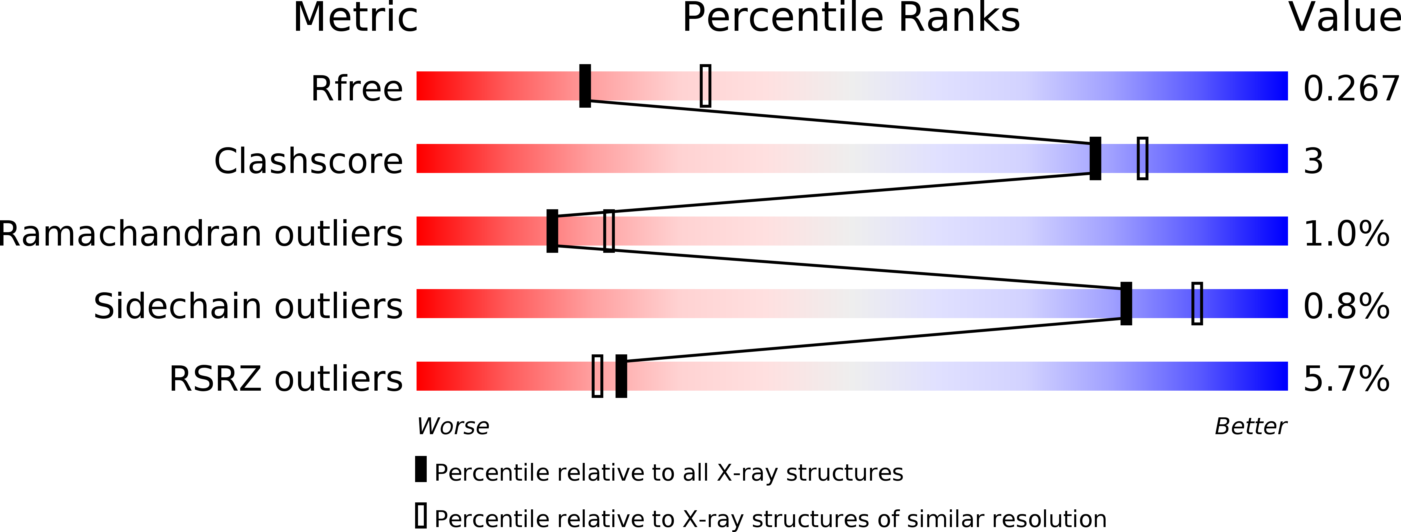
Resolution:
2.64 Å
R-Value Free:
0.26
R-Value Work:
0.19
R-Value Observed:
0.19
Space Group:
P 21 21 21
Macromolecular Entities
Polymer Type:polypeptide(L)
Molecule:Enoyl-CoA delta isomerase 2, mitochondrial
Gene (Uniprot):ECI2
Chain IDs:A, B, C
Chain Length:276
Number of Molecules:3
Biological Source:Homo sapiens
Primary Citation
Human Delta (3) , Delta (2) -enoyl-CoA isomerase, type 2: a structural enzymology study on the catalytic role of its ACBP domain and helix-10.
Febs J. 282 746 768 (2015)
PMID: 25515061 DOI: 10.1111/febs.13179

Abstact

The catalytic domain of the trimeric human Δ(3),Δ(2)-enoyl-CoA isomerase, type 2 (HsECI2), has the typical crotonase fold. In the active site of this fold two main chain NH groups form an oxyanion hole for binding the thioester oxygen of the 3E- or 3Z-enoyl-CoA substrate molecules. A catalytic glutamate is essential for the proton transfer between the substrate C2 and C4 atoms for forming the product 2E-enoyl-CoA, which is a key intermediate in the β-oxidation pathway. The active site is covered by the C-terminal helix-10. In HsECI2, the isomerase domain is extended at its N terminus by an acyl-CoA binding protein (ACBP) domain. Small angle X-ray scattering analysis of HsECI2 shows that the ACBP domain protrudes out of the central isomerase trimer. X-ray crystallography of the isomerase domain trimer identifies the active site geometry. A tunnel, shaped by loop-2 and extending from the catalytic site to bulk solvent, suggests a likely mode of binding of the fatty acyl chains. Calorimetry data show that the separately expressed ACBP and isomerase domains bind tightly to fatty acyl-CoA molecules. The truncated isomerase variant (without ACBP domain) has significant enoyl-CoA isomerase activity; however, the full-length isomerase is more efficient. Structural enzymological studies of helix-10 variants show the importance of this helix for efficient catalysis. Its hydrophobic side chains, together with residues from loop-2 and loop-4, complete a hydrophobic cluster that covers the active site, thereby fixing the thioester moiety in a mode of binding competent for efficient catalysis.

Legend

Protein

Chemical

Disease

Primary Citation of related structures