Deposition Date
2014-07-15
Release Date
2014-11-26
Last Version Date
2024-11-06
Entry Detail
PDB ID:
4U14
Keywords:
Title:
Structure of the M3 muscarinic acetylcholine receptor bound to the antagonist tiotropium crystallized with disulfide-stabilized T4 lysozyme (dsT4L)
Biological Source:
Source Organism(s):
Rattus norvegicus (Taxon ID: 10116)
Enterobacteria phage T4 (Taxon ID: 10665)
Enterobacteria phage T4 (Taxon ID: 10665)
Expression System(s):
Method Details:
Experimental Method:
Resolution:
3.57 Å
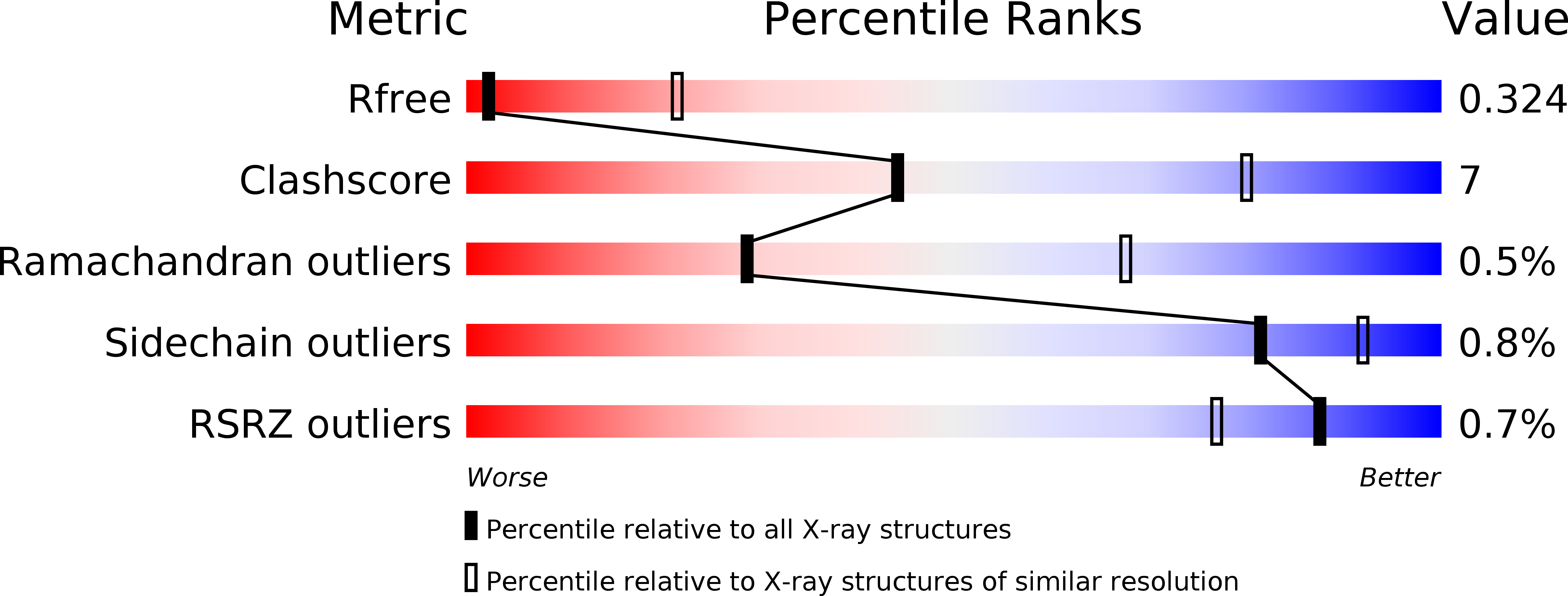
R-Value Free:
0.32
R-Value Work:
0.27
R-Value Observed:
0.27
Space Group:
P 41 21 2