Deposition Date
2014-06-29
Release Date
2014-11-12
Last Version Date
2023-12-20
Entry Detail
PDB ID:
4TW1
Keywords:
Title:
Crystal structure of the octameric pore complex of the Staphylococcus aureus Bi-component Toxin LukGH
Biological Source:
Source Organism(s):
Staphylococcus aureus (Taxon ID: 451516)
Expression System(s):
Method Details:
Experimental Method:
Resolution:
2.80 Å
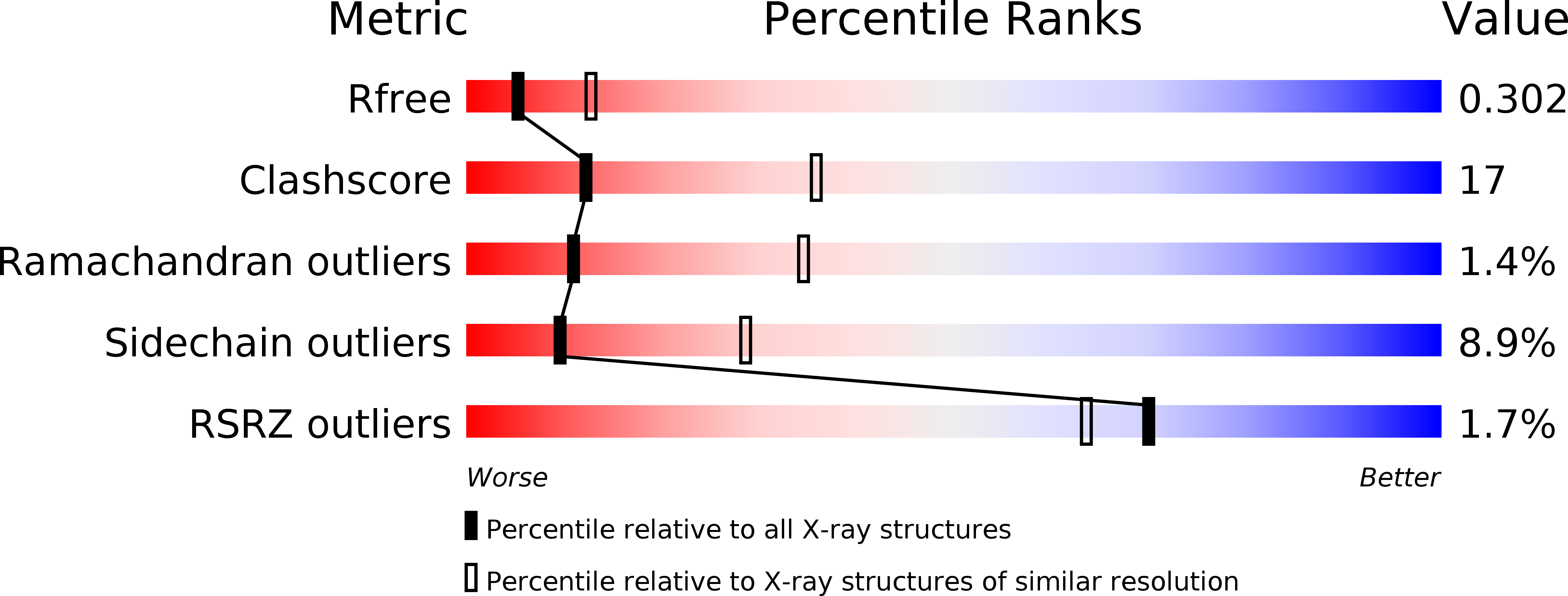
R-Value Free:
0.29
R-Value Work:
0.23
R-Value Observed:
0.23
Space Group:
P 1 21 1