4TVW image
Entry Detail
PDB ID:
4TVW
Keywords:
Title:
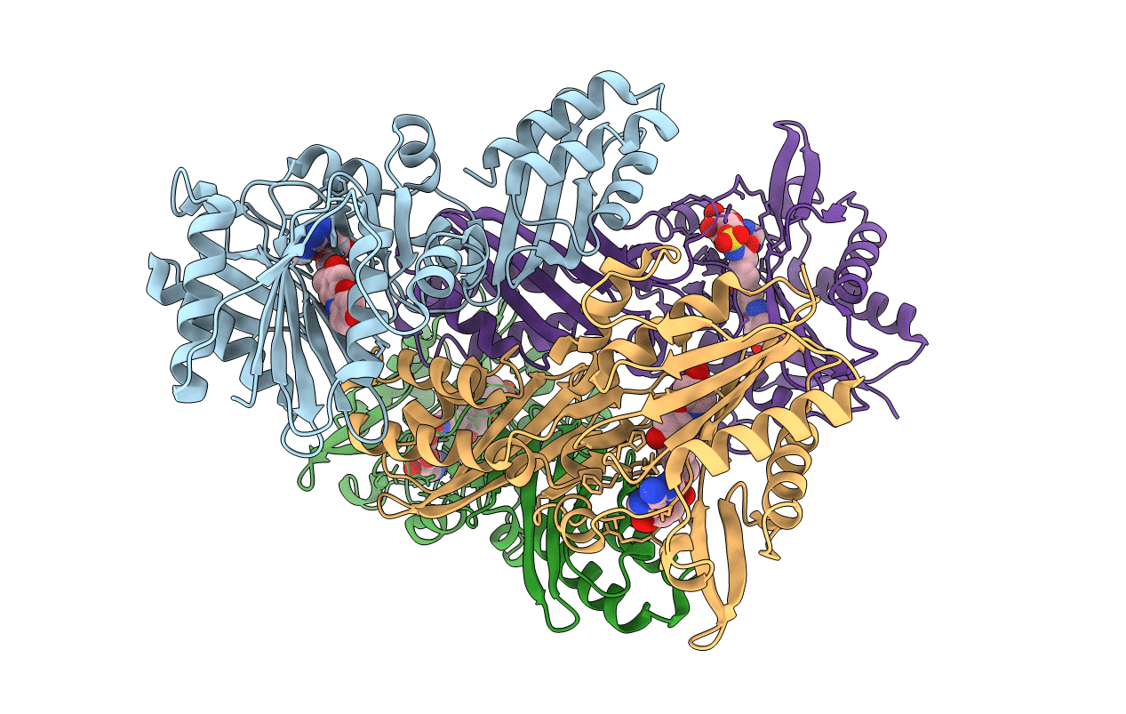
Resorufin ligase with bound resorufin-AMP analog
Biological Source:
Source Organism:
Host Organism:
PDB Version:
Deposition Date:
2014-06-28
Release Date:
2014-10-22
Method Details:
Experimental Method:
Resolution:
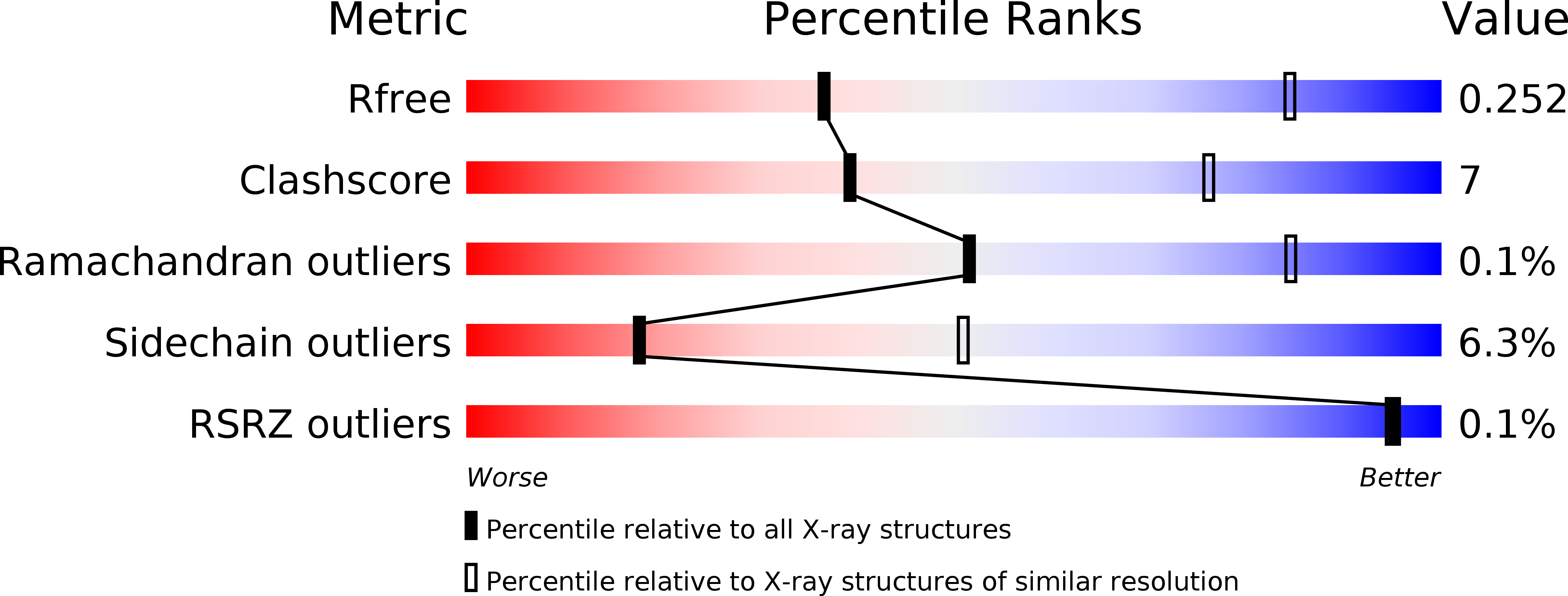
3.51 Å
R-Value Free:
0.24
R-Value Work:
0.20
R-Value Observed:
0.20
Space Group:
P 1 21 1
Macromolecular Entities
Polymer Type:polypeptide(L)
Description:Lipoate-protein ligase A
Mutations:E20A, F147A, H149G
Chain IDs:A, B, C, D
Chain Length:341
Number of Molecules:4
Biological Source:Escherichia coli
Ligand Molecules
Primary Citation
Computational design of a red fluorophore ligase for site-specific protein labeling in living cells.
Proc.Natl.Acad.Sci.USA 111 E4551 E4559 (2014)
PMID: 25313043 DOI: 10.1073/pnas.1404736111

Abstact

Chemical fluorophores offer tremendous size and photophysical advantages over fluorescent proteins but are much more challenging to target to specific cellular proteins. Here, we used Rosetta-based computation to design a fluorophore ligase that accepts the red dye resorufin, starting from Escherichia coli lipoic acid ligase. X-ray crystallography showed that the design closely matched the experimental structure. Resorufin ligase catalyzed the site-specific and covalent attachment of resorufin to various cellular proteins genetically fused to a 13-aa recognition peptide in multiple mammalian cell lines and in primary cultured neurons. We used resorufin ligase to perform superresolution imaging of the intermediate filament protein vimentin by stimulated emission depletion and electron microscopies. This work illustrates the power of Rosetta for major redesign of enzyme specificity and introduces a tool for minimally invasive, highly specific imaging of cellular proteins by both conventional and superresolution microscopies.

Legend

Protein

Chemical

Disease

Primary Citation of related structures