4TV8 image
Deposition Date 2014-06-26
Release Date 2014-08-27
Last Version Date 2023-12-20
Entry Detail
PDB ID:
4TV8
Keywords:
Title:
Tubulin-Maytansine complex
Biological Source:
Source Organism:
Rattus norvegicus (Taxon ID: 10116)
Gallus gallus (Taxon ID: 9031)
Bos taurus (Taxon ID: 9913)
Host Organism:
Method Details:
Experimental Method:
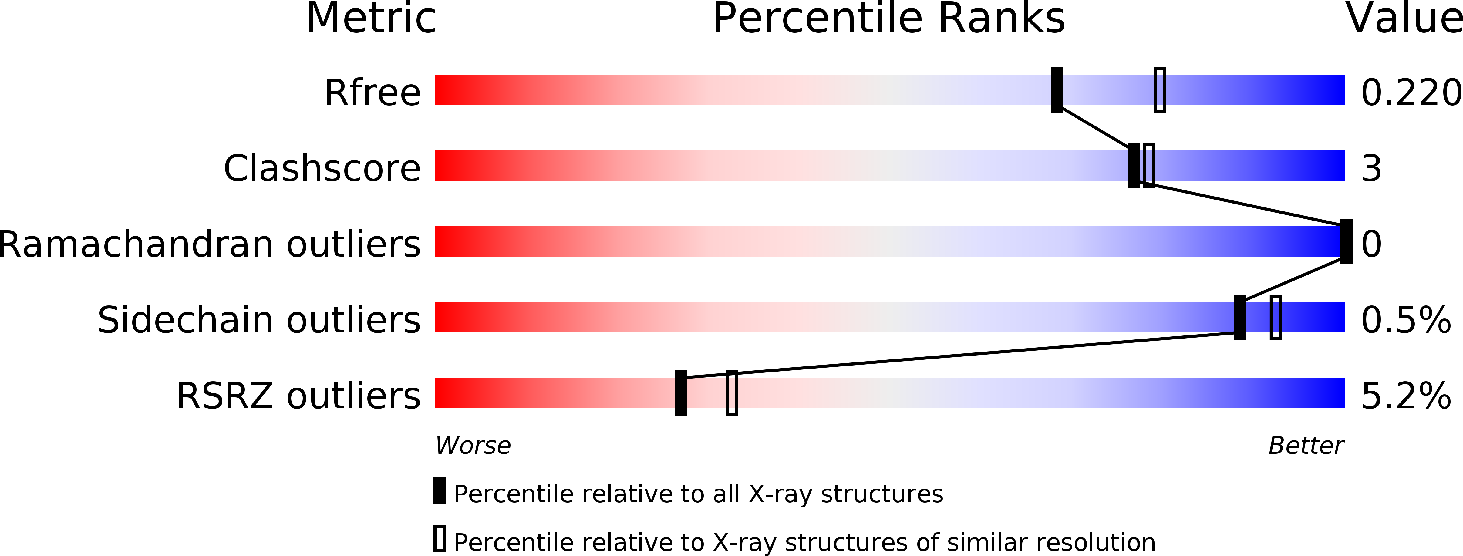
Resolution:
2.10 Å
R-Value Free:
0.21
R-Value Work:
0.18
R-Value Observed:
0.19
Space Group:
P 21 21 21
Macromolecular Entities
Structures with similar UniProt ID
Protein Blast
Polymer Type:polypeptide(L)
Molecule:Tubulin alpha-1B chain
Chain IDs:A, C
Chain Length:451
Number of Molecules:2
Biological Source:Bos taurus
Structures with similar UniProt ID
Protein Blast
Polymer Type:polypeptide(L)
Molecule:Tubulin beta-2B chain
Gene (Uniprot):TUBB2B
Chain IDs:B, D
Chain Length:445
Number of Molecules:2
Biological Source:Bos taurus
Structures with similar UniProt ID
Protein Blast
Polymer Type:polypeptide(L)
Molecule:Stathmin-4
Gene (Uniprot):Stmn4
Chain IDs:E
Chain Length:143
Number of Molecules:1
Biological Source:Rattus norvegicus
Polymer Type:polypeptide(L)
Molecule:Tubulin-Tyrosine Ligase
Chain IDs:F
Chain Length:384
Number of Molecules:1
Biological Source:Gallus gallus
Primary Citation
A new tubulin-binding site and pharmacophore for microtubule-destabilizing anticancer drugs.
Proc.Natl.Acad.Sci.USA 111 13817 13821 (2014)
PMID: 25114240 DOI: 10.1073/pnas.1408124111

Abstact

The recent success of antibody-drug conjugates (ADCs) in the treatment of cancer has led to a revived interest in microtubule-destabilizing agents. Here, we determined the high-resolution crystal structure of the complex between tubulin and maytansine, which is part of an ADC that is approved by the US Food and Drug Administration (FDA) for the treatment of advanced breast cancer. We found that the drug binds to a site on β-tubulin that is distinct from the vinca domain and that blocks the formation of longitudinal tubulin interactions in microtubules. We also solved crystal structures of tubulin in complex with both a variant of rhizoxin and the phase 1 drug PM060184. Consistent with biochemical and mutagenesis data, we found that the two compounds bound to the same site as maytansine and that the structures revealed a common pharmacophore for the three ligands. Our results delineate a distinct molecular mechanism of action for the inhibition of microtubule assembly by clinically relevant agents. They further provide a structural basis for the rational design of potent microtubule-destabilizing agents, thus opening opportunities for the development of next-generation ADCs for the treatment of cancer.

Legend

Protein

Chemical

Disease

Primary Citation of related structures
Feedback Form
Name
Email
Institute
Feedback