4TV5 image
Deposition Date 2014-06-25
Release Date 2014-10-29
Last Version Date 2023-12-27
Entry Detail
PDB ID:
4TV5
Keywords:
Title:
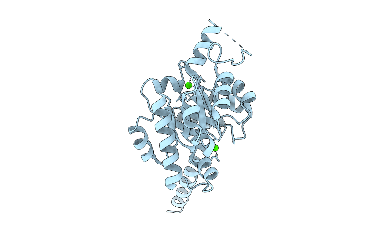
Crystal Structure of Citrate Synthase SbnG
Biological Source:
Source Organism:
Host Organism:
Method Details:
Experimental Method:
Resolution:
1.85 Å
R-Value Free:
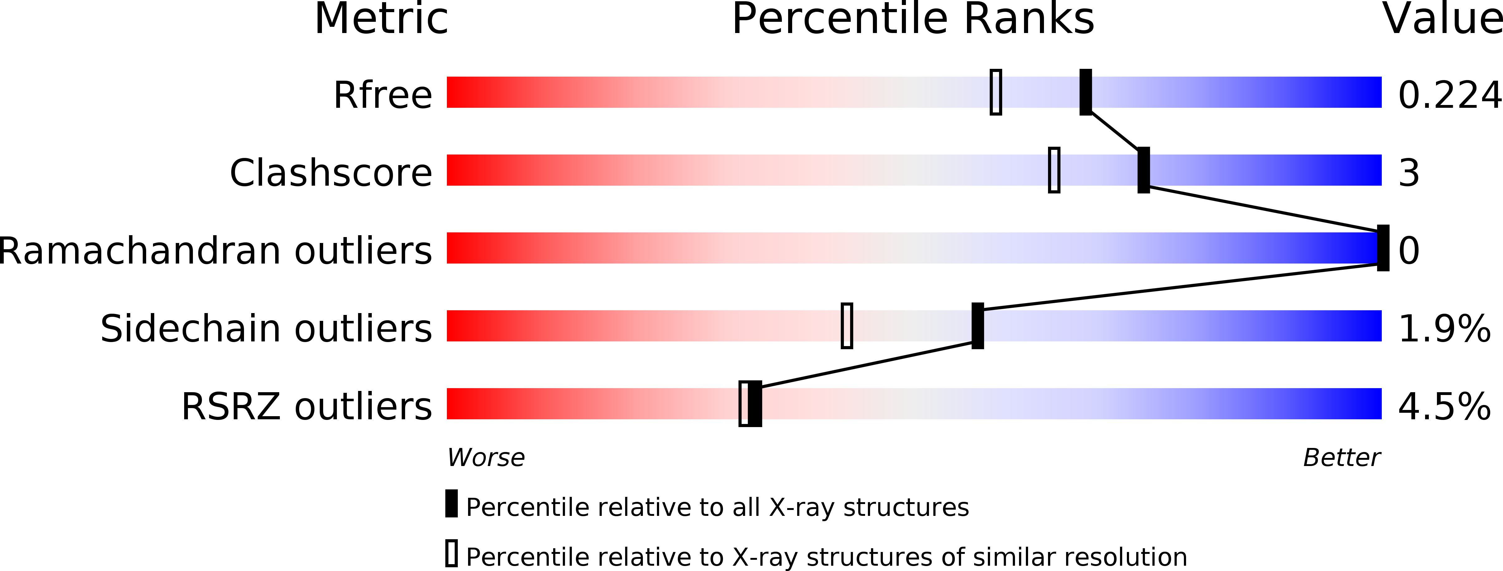
0.21
R-Value Work:
0.19
R-Value Observed:
0.19
Space Group:
P 3 1 2
Macromolecular Entities
Polymer Type:polypeptide(L)
Molecule:2-dehydro-3-deoxyglucarate aldolase
Chain IDs:A
Chain Length:259
Number of Molecules:1
Biological Source:Staphylococcus aureus
Ligand Molecules
Primary Citation
SbnG, a Citrate Synthase in Staphylococcus aureus: A NEW FOLD ON AN OLD ENZYME.
J.Biol.Chem. 289 33797 33807 (2014)
PMID: 25336653 DOI: 10.1074/jbc.M114.603175

Abstact

In response to iron deprivation, Staphylococcus aureus produces staphyloferrin B, a citrate-containing siderophore that delivers iron back to the cell. This bacterium also possesses a second citrate synthase, SbnG, that is necessary for supplying citrate to the staphyloferrin B biosynthetic pathway. We present the structure of SbnG bound to the inhibitor calcium and an active site variant in complex with oxaloacetate. The overall fold of SbnG is structurally distinct from TCA cycle citrate synthases yet similar to metal-dependent class II aldolases. Phylogenetic analyses revealed that SbnG forms a separate clade with homologs from other siderophore biosynthetic gene clusters and is representative of a metal-independent subgroup in the phosphoenolpyruvate/pyruvate domain superfamily. A structural superposition of the SbnG active site to TCA cycle citrate synthases and site-directed mutagenesis suggests a case for convergent evolution toward a conserved catalytic mechanism for citrate production.

Legend

Protein

Chemical

Disease

Primary Citation of related structures