4TUO image
Deposition Date 2014-06-24
Release Date 2015-07-15
Last Version Date 2024-11-06
Entry Detail
PDB ID:
4TUO
Keywords:
Title:
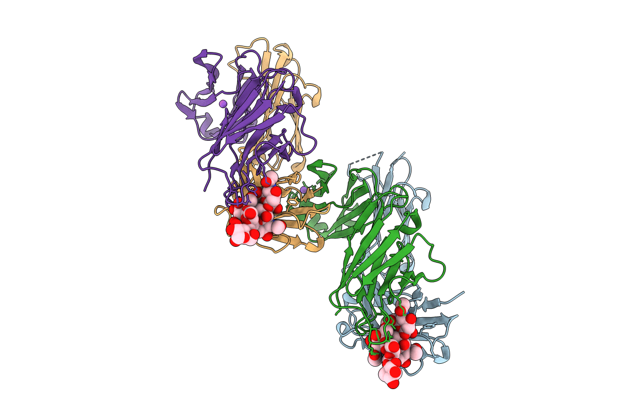
Crystal structure of monoclonal antibody against neuroblastoma associated antigen.
Biological Source:
Source Organism:
Mus musculus (Taxon ID: 10090)
Host Organism:
Method Details:
Experimental Method:
Resolution:
1.55 Å
R-Value Free:
0.20
R-Value Work:
0.16
R-Value Observed:
0.16
Space Group:
P 1
Macromolecular Entities
Polymer Type:polypeptide(L)
Molecule:Heavy chain of monoclonal antibody against neuroblastoma associated antigen
Chain IDs:A, C
Chain Length:214
Number of Molecules:2
Biological Source:Mus musculus
Polymer Type:polypeptide(L)
Molecule:Light chain of monoclonal antibody against neuroblastoma associated antigen
Chain IDs:B, D
Chain Length:220
Number of Molecules:2
Biological Source:Mus musculus
Ligand Molecules
Primary Citation
Structural Basis of GD2 Ganglioside and Mimetic Peptide Recognition by 14G2a Antibody.
Mol.Cell Proteomics 14 2577 2590 (2015)
PMID: 26179345 DOI: 10.1074/mcp.M115.052720

Abstact

Monoclonal antibodies targeting GD2 ganglioside (GD2) have recently been approved for the treatment of high risk neuroblastoma and are extensively evaluated in clinics in other indications. This study illustrates how a therapeutic antibody distinguishes between different types of gangliosides present on normal and cancer cells and informs how synthetic peptides can imitate ganglioside in its binding to the antibody. Using high resolution crystal structures we demonstrate that the ganglioside recognition by a model antibody (14G2a) is based primarily on an extended network of direct and water molecule mediated hydrogen bonds. Comparison of the GD2-Fab structure with that of a ligand free antibody reveals an induced fit mechanism of ligand binding. These conclusions are validated by directed mutagenesis and allowed structure guided generation of antibody variant with improved affinity toward GD2. Contrary to the carbohydrate, both evaluated mimetic peptides utilize a "key and lock" interaction mechanism complementing the surface of the antibody binding groove exactly as found in the empty structure. The interaction of both peptides with the Fab relies considerably on hydrophobic contacts however, the detailed connections differ significantly between the peptides. As such, the evaluated peptide carbohydrate mimicry is defined primarily in a functional and not in structural manner.

Legend

Protein

Chemical

Disease

Primary Citation of related structures
Feedback Form
Name
Email
Institute
Feedback