
Deposition Date
2014-06-19
Release Date
2014-08-06
Last Version Date
2023-12-20
Entry Detail
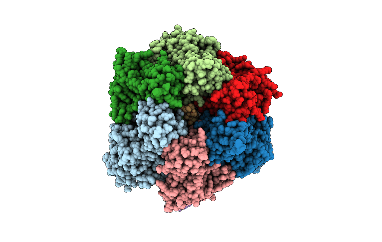
PDB ID:
4TT3
Keywords:
Title:
The Pathway of Binding of the Intrinsically Disordered Mitochondrial Inhibitor Protein to F1-ATPase
Biological Source:
Source Organism(s):
Bos taurus (Taxon ID: 9913)
Expression System(s):
Method Details:
Experimental Method:
Resolution:
3.21 Å
R-Value Free:
0.28
R-Value Work:
0.24
R-Value Observed:
0.24
Space Group:
P 21 21 21
