4TRK image
Deposition Date 2014-06-17
Release Date 2014-11-19
Last Version Date 2023-12-27
Entry Detail
PDB ID:
4TRK
Title:
Structure of C. elegans HIM-3
Biological Source:
Source Organism:
Host Organism:
Method Details:
Experimental Method:
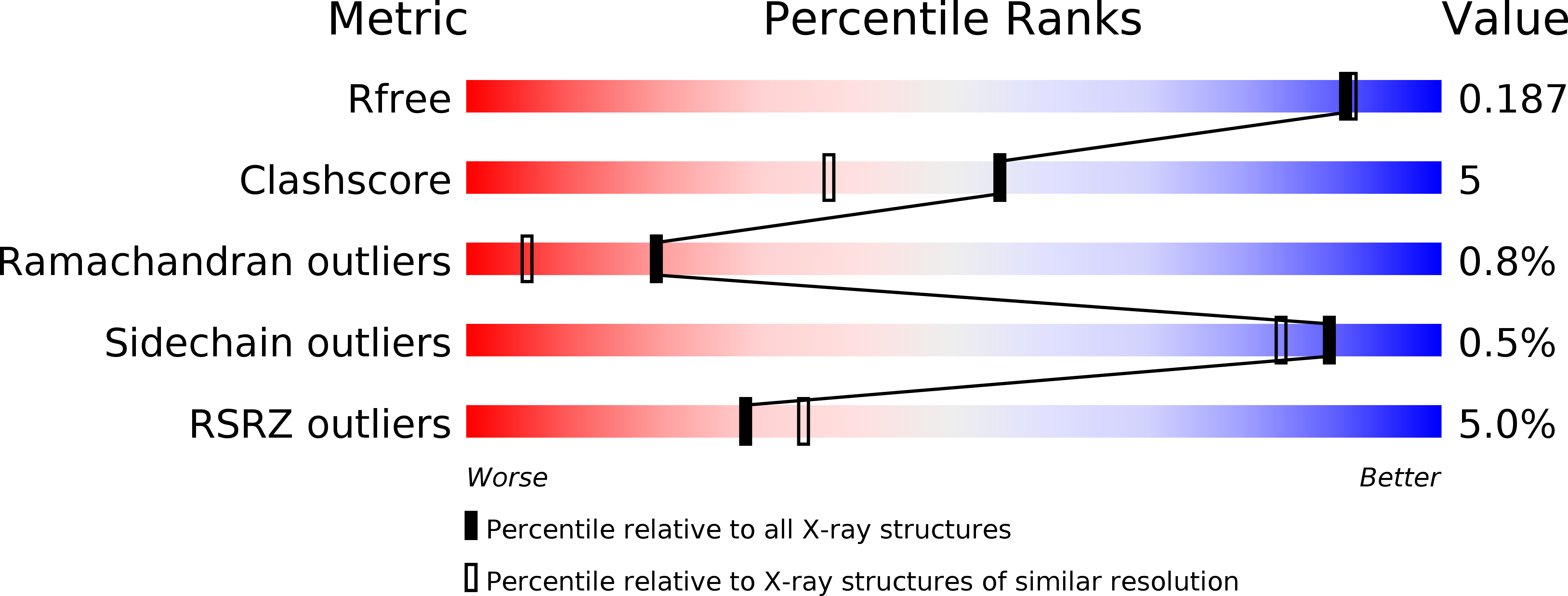
Resolution:
1.75 Å
R-Value Free:
0.18
R-Value Work:
0.15
R-Value Observed:
0.15
Space Group:
P 61
Macromolecular Entities
Polymer Type:polypeptide(L)
Molecule:C. elegans HIM-3
Gene (Uniprot):him-3
Chain IDs:A
Chain Length:291
Number of Molecules:1
Biological Source:Caenorhabditis elegans
Primary Citation
The Chromosome Axis Controls Meiotic Events through a Hierarchical Assembly of HORMA Domain Proteins.
Dev.Cell 31 487 502 (2014)
PMID: 25446517 DOI: 10.1016/j.devcel.2014.09.013

Abstact

Proteins of the HORMA domain family play central, but poorly understood, roles in chromosome organization and dynamics during meiosis. In Caenorhabditis elegans, four such proteins (HIM-3, HTP-1, HTP-2, and HTP-3) have distinct but overlapping functions. Through combined biochemical, structural, and in vivo analysis, we find that these proteins form hierarchical complexes through binding of their HORMA domains to cognate peptides within their partners' C-terminal tails, analogous to the "safety belt" binding mechanism of Mad2. These interactions are critical for recruitment of HIM-3, HTP-1, and HTP-2 to chromosome axes. HTP-3, in addition to recruiting the other HORMA domain proteins to the axis, plays an independent role in sister chromatid cohesion and double-strand break formation. Finally, we find that mammalian HORMAD1 binds a motif found both at its own C terminus and at that of HORMAD2, indicating that this mode of intermolecular association is a conserved feature of meiotic chromosome structure in eukaryotes.

Legend

Protein

Chemical

Disease

Primary Citation of related structures