4TR6 image
Entry Detail
PDB ID:
4TR6
Title:
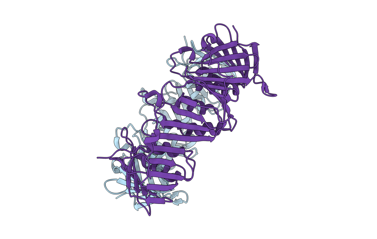
Crystal structure of DNA polymerase sliding clamp from Bacillus subtilis
Biological Source:
Source Organism:
Host Organism:
PDB Version:
Deposition Date:
2014-06-14
Release Date:
2014-09-10
Method Details:
Experimental Method:
Resolution:
1.50 Å
R-Value Free:
0.20
R-Value Work:
0.17
R-Value Observed:
0.17
Space Group:
P 1
Macromolecular Entities
Polymer Type:polypeptide(L)
Description:DNA polymerase III subunit beta
Chain IDs:A, B
Chain Length:380
Number of Molecules:2
Biological Source:Bacillus subtilis
Ligand Molecules
Primary Citation
Differential Modes of Peptide Binding onto Replicative Sliding Clamps from Various Bacterial Origins.
J.Med.Chem. 57 7565 7576 (2014)
PMID: 25170813 DOI: 10.1021/jm500467a

Abstact

Bacterial sliding clamps are molecular hubs that interact with many proteins involved in DNA metabolism through their binding, via a conserved peptidic sequence, into a universally conserved pocket. This interacting pocket is acknowledged as a potential molecular target for the development of new antibiotics. We previously designed short peptides with an improved affinity for the Escherichia coli binding pocket. Here we show that these peptides differentially interact with other bacterial clamps, despite the fact that all pockets are structurally similar. Thermodynamic and modeling analyses of the interactions differentiate between two categories of clamps: group I clamps interact efficiently with our designed peptides and assemble the Escherichia coli and related orthologs clamps, whereas group II clamps poorly interact with the same peptides and include Bacillus subtilis and other Gram-positive clamps. These studies also suggest that the peptide binding process could occur via different mechanisms, which depend on the type of clamp.

Legend

Protein

Chemical

Disease

Primary Citation of related structures