Deposition Date
2014-06-11
Release Date
2014-09-24
Last Version Date
2024-11-20
Entry Detail
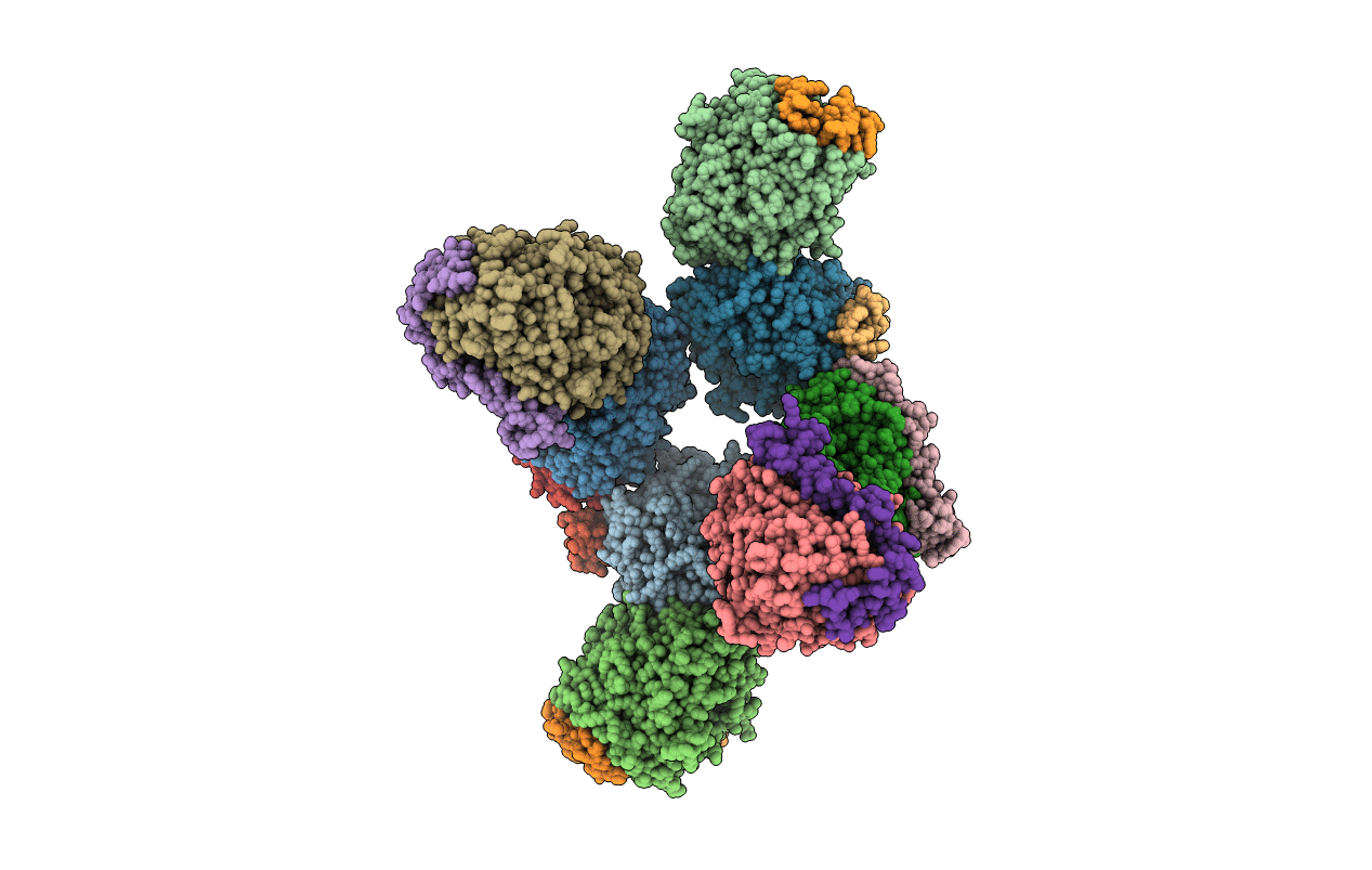
PDB ID:
4TQO
Keywords:
Title:
The crystal structure of methanol dehydrogenase from Methylococcus capsulatus (Bath)
Biological Source:
Source Organism(s):
Methylococcus capsulatus (Taxon ID: 243233)
Method Details:
Experimental Method:
Resolution:
2.57 Å
R-Value Free:
0.20
R-Value Work:
0.15
R-Value Observed:
0.16
Space Group:
P 21 21 21