
Deposition Date
2014-06-10
Release Date
2014-07-16
Last Version Date
2023-09-27
Entry Detail
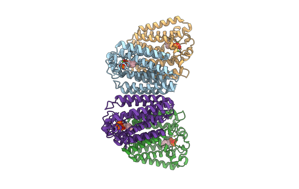
PDB ID:
4TQ4
Keywords:
Title:
Structure of a UbiA homolog from Archaeoglobus fulgidus bound to DMAPP and Mg2+
Biological Source:
Source Organism(s):
Archaeoglobus fulgidus (Taxon ID: 224325)
Expression System(s):
Method Details:
Experimental Method:
Resolution:
2.50 Å
R-Value Free:
0.25
R-Value Work:
0.21
R-Value Observed:
0.21
Space Group:
P 1 21 1
