Deposition Date
2014-06-05
Release Date
2015-02-11
Last Version Date
2023-12-27
Entry Detail
PDB ID:
4TOB
Keywords:
Title:
1.95A resolution structure of BfrB (Q151L) from Pseudomonas aeruginosa
Biological Source:
Source Organism(s):
Pseudomonas aeruginosa (Taxon ID: 208964)
Expression System(s):
Method Details:
Experimental Method:
Resolution:
1.95 Å
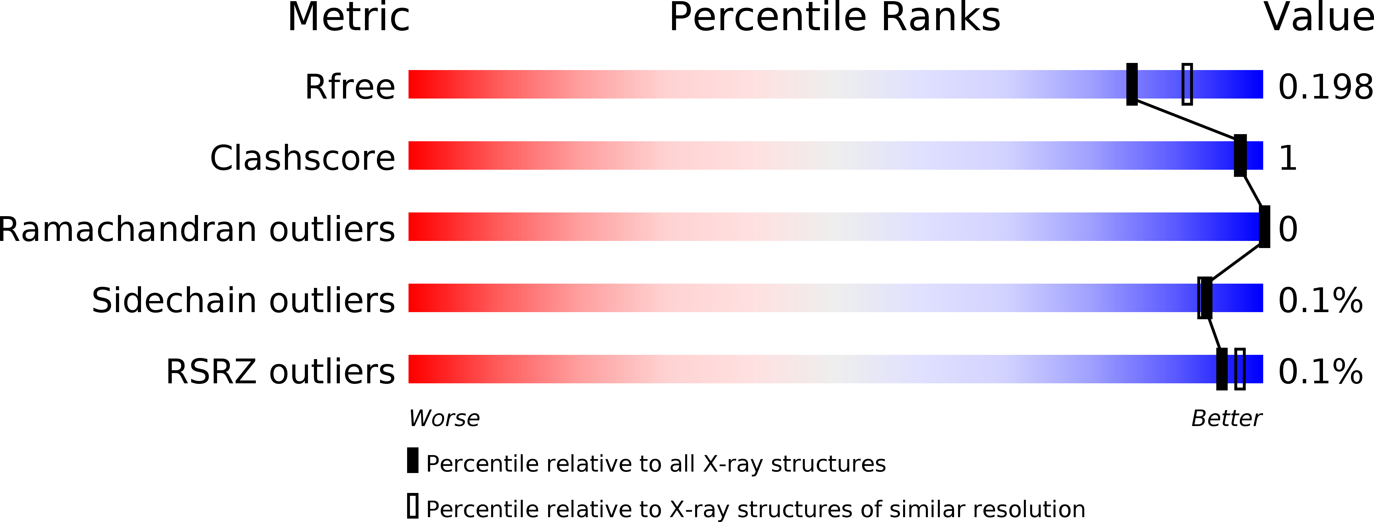
R-Value Free:
0.18
R-Value Work:
0.15
R-Value Observed:
0.15
Space Group:
P 21 21 21