
Deposition Date
2014-06-05
Release Date
2014-07-30
Last Version Date
2023-09-27
Entry Detail
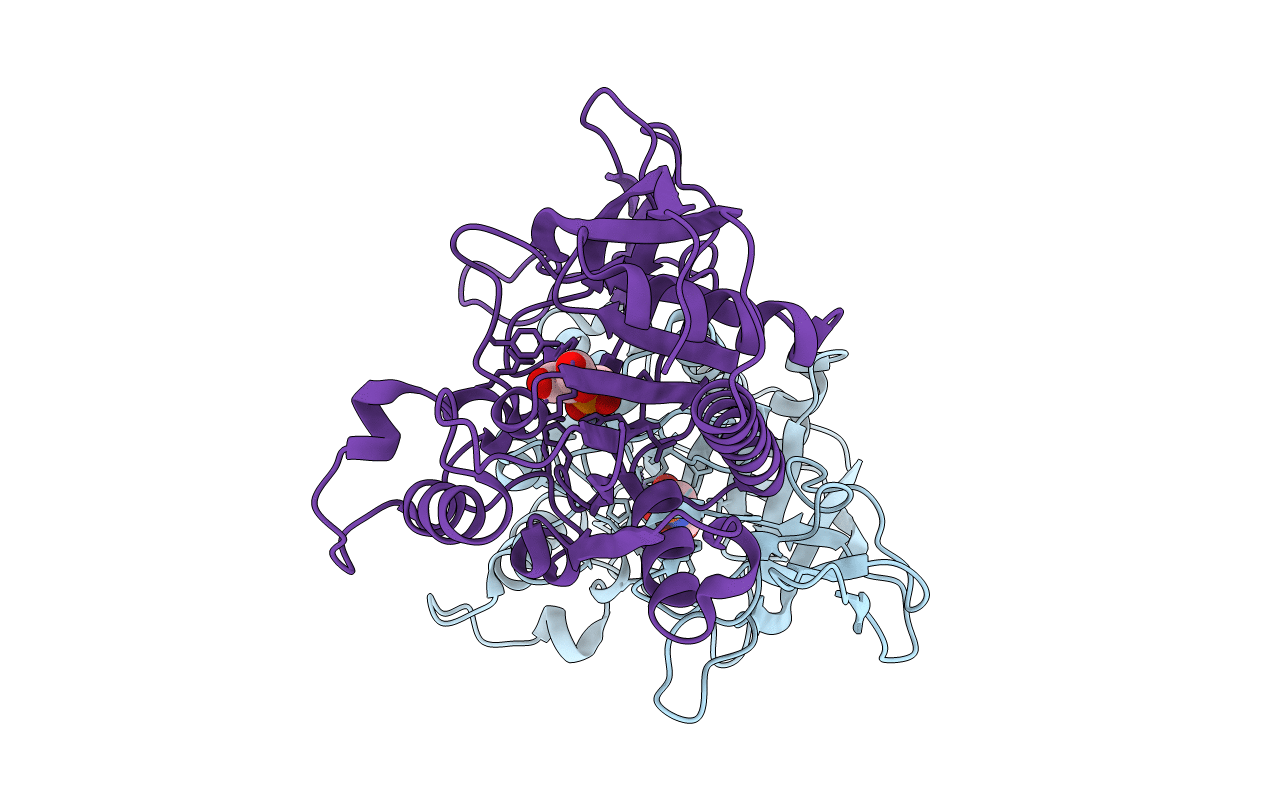
PDB ID:
4TNU
Keywords:
Title:
Human brain aspartoacylase mutant Y231C complex with intermediate analog (N-phosphonomethyl-L-aspartate)
Biological Source:
Source Organism(s):
Homo sapiens (Taxon ID: 9606)
Expression System(s):
Method Details:
Experimental Method:
Resolution:
2.90 Å
R-Value Free:
0.23
R-Value Work:
0.19
R-Value Observed:
0.19
Space Group:
P 42 21 2
