4TMB image
Deposition Date 2014-05-31
Release Date 2015-02-11
Last Version Date 2023-11-08
Entry Detail
PDB ID:
4TMB
Keywords:
Title:
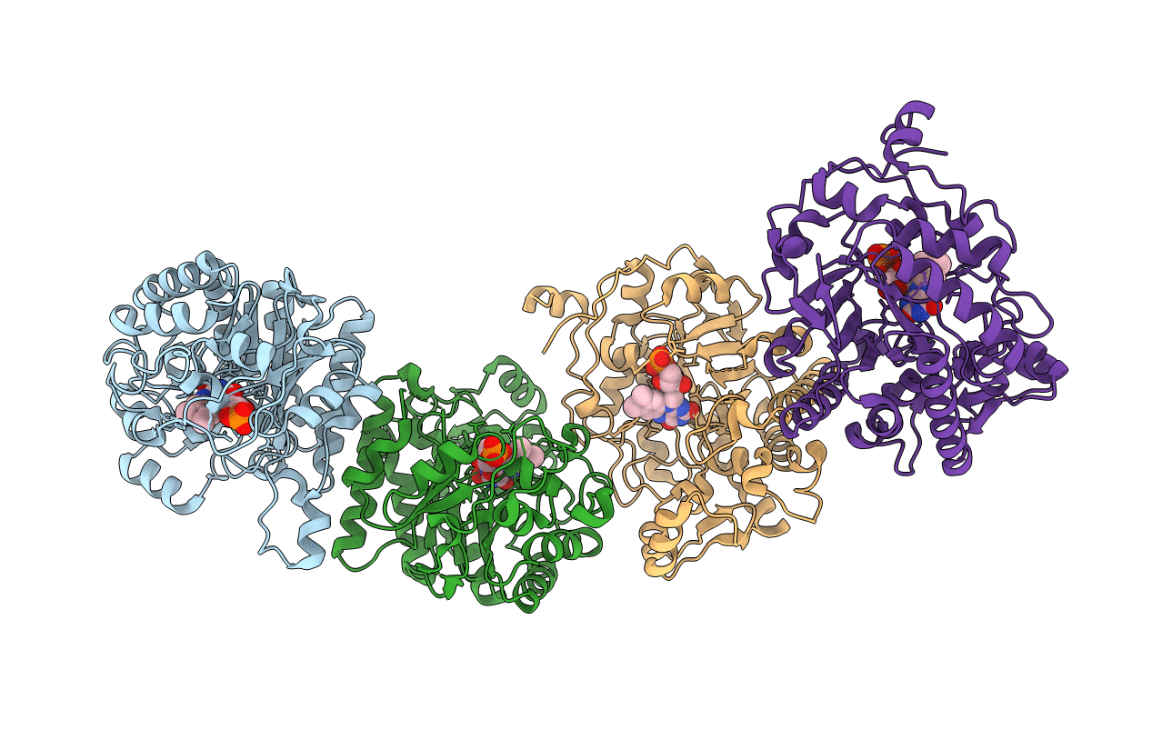
CRYSTAL STRUCTURE of OLD YELLOW ENZYME from CANDIDA MACEDONIENSIS AKU4588
Biological Source:
Source Organism:
Host Organism:
Method Details:
Experimental Method:
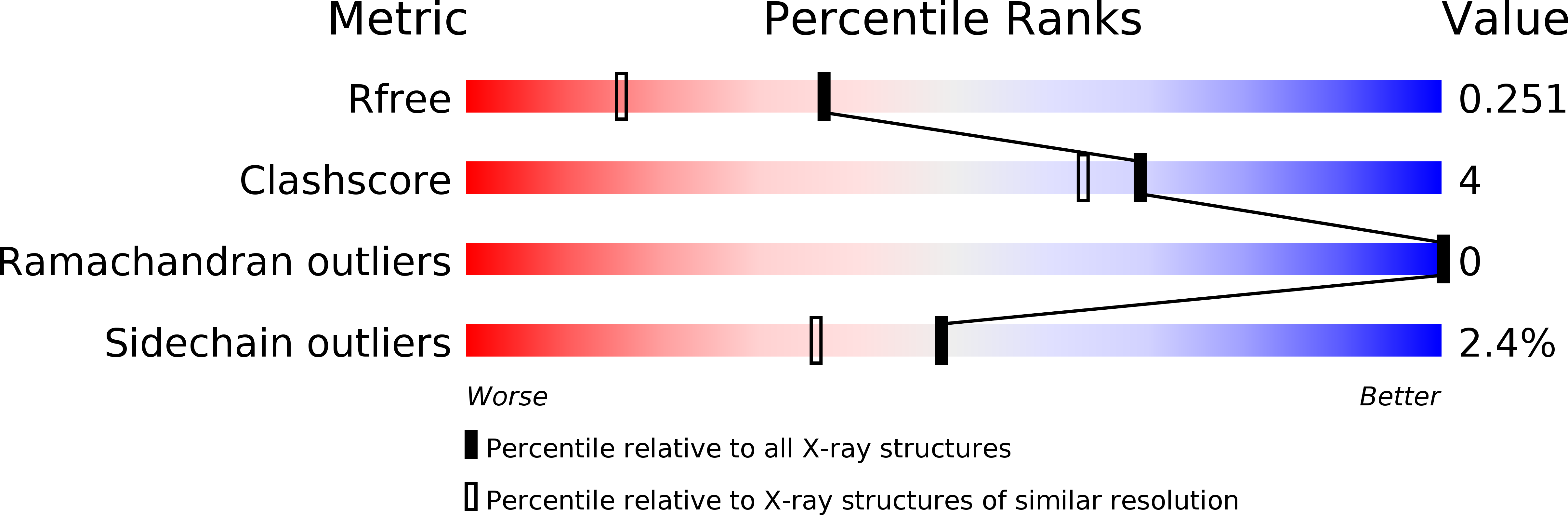
Resolution:
1.80 Å
R-Value Free:
0.21
R-Value Work:
0.17
R-Value Observed:
0.17
Space Group:
C 1 2 1
Macromolecular Entities
Structures with similar UniProt ID
Protein Blast
Polymer Type:polypeptide(L)
Molecule:Old yellow enzyme
Gene (Uniprot):oye
Chain IDs:A, B, C, D
Chain Length:403
Number of Molecules:4
Biological Source:Kluyveromyces marxianus
Ligand Molecules
Primary Citation
An Engineered Old Yellow Enzyme that Enables Efficient Synthesis of (4R,6R)-Actinol in a One-Pot Reduction System
Chembiochem 16 440 445 (2015)
PMID: 25639703 DOI: 10.1002/cbic.201402555

Abstact

(4R,6R)-Actinol can be stereo-selectively synthesized from ketoisophorone by a two-step conversion using a mixture of two enzymes: Candida macedoniensis old yellow enzyme (CmOYE) and Corynebacterium aquaticum (6R)-levodione reductase. However, (4S)-phorenol, an intermediate, accumulates because of the limited substrate range of CmOYE. To address this issue, we solved crystal structures of CmOYE in the presence and absence of a substrate analogue p-HBA, and introduced point mutations into the substrate-recognition loop. The most effective mutant (P295G) showed two- and 12-fold higher catalytic activities toward ketoisophorone and (4S)-phorenol, respectively, than the wild-type, and improved the yield of the two-step conversion from 67.2 to 90.1%. Our results demonstrate that the substrate range of an enzyme can be changed by introducing mutation(s) into a substrate-recognition loop. This method can be applied to the development of other favorable OYEs with different substrate preferences.

Legend

Protein

Chemical

Disease

Primary Citation of related structures
Feedback Form
Name
Email
Institute
Feedback