4TLL image
Deposition Date 2014-05-30
Release Date 2014-07-02
Last Version Date 2023-12-27
Entry Detail
PDB ID:
4TLL
Title:
Crystal structure of GluN1/GluN2B NMDA receptor, structure 1
Biological Source:
Source Organism:
Xenopus laevis (Taxon ID: 8355)
Host Organism:
Method Details:
Experimental Method:
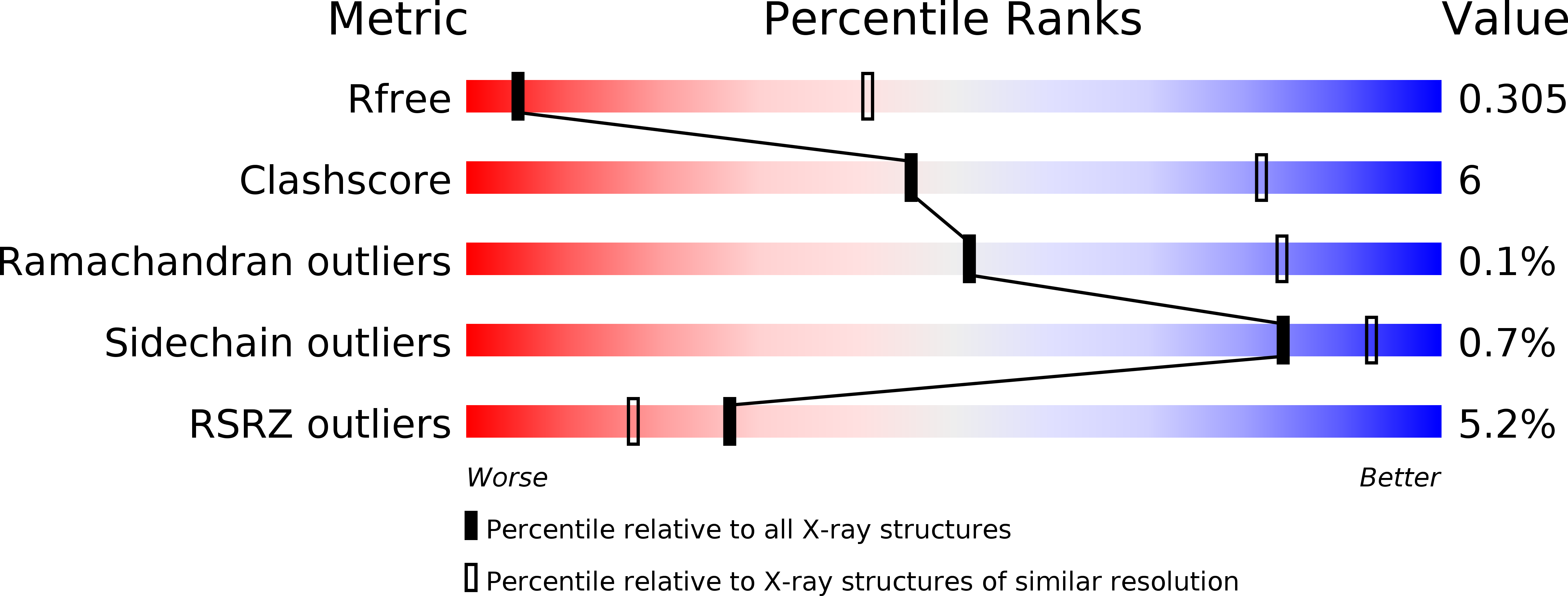
Resolution:
3.59 Å
R-Value Free:
0.30
R-Value Work:
0.26
R-Value Observed:
0.26
Space Group:
C 1 2 1
Macromolecular Entities
Polymer Type:polypeptide(L)
Molecule:receptor subunit GluN1
Mutations:C22A, K51F, K52F, N300Q, N350Q, N368D, N440D, N469D, K493A, K494A, E495A, G610R, I617L, D656R, N769E, Del(K588-E595), Replacement of 845-853 with SRAEAKRMK
Chain IDs:A, C
Chain Length:823
Number of Molecules:2
Biological Source:Xenopus laevis
Polymer Type:polypeptide(L)
Molecule:receptor subunit GluN2B
Gene (Uniprot):grin2b
Mutations:M20S, G21R, C22A, A64E, N69Q, K216C, N343D, T490V, C585A, E654R, E655R, V615L, Del(K382-V385), Del(R584-G593), Replacement of 854-864 with YKSRAEAKRMK
Chain IDs:B, D
Chain Length:824
Number of Molecules:2
Biological Source:Xenopus laevis
Primary Citation
NMDA receptor structures reveal subunit arrangement and pore architecture.
Nature 511 191 197 (2014)
PMID: 25008524 DOI: 10.1038/nature13548

Abstact

N-methyl-d-aspartate (NMDA) receptors are Hebbian-like coincidence detectors, requiring binding of glycine and glutamate in combination with the relief of voltage-dependent magnesium block to open an ion conductive pore across the membrane bilayer. Despite the importance of the NMDA receptor in the development and function of the brain, a molecular structure of an intact receptor has remained elusive. Here we present X-ray crystal structures of the Xenopus laevis GluN1-GluN2B NMDA receptor with the allosteric inhibitor, Ro25-6981, partial agonists and the ion channel blocker, MK-801. Receptor subunits are arranged in a 1-2-1-2 fashion, demonstrating extensive interactions between the amino-terminal and ligand-binding domains. The transmembrane domains harbour a closed-blocked ion channel, a pyramidal central vestibule lined by residues implicated in binding ion channel blockers and magnesium, and a ∼twofold symmetric arrangement of ion channel pore loops. These structures provide new insights into the architecture, allosteric coupling and ion channel function of NMDA receptors.

Legend

Protein

Chemical

Disease

Primary Citation of related structures