4TL6 image
Entry Detail
PDB ID:
4TL6
Keywords:
Title:
Crystal structure of N-terminal domain of KaiC
Biological Source:
PDB Version:
Deposition Date:
2014-05-29
Release Date:
2015-07-01
Method Details:
Experimental Method:
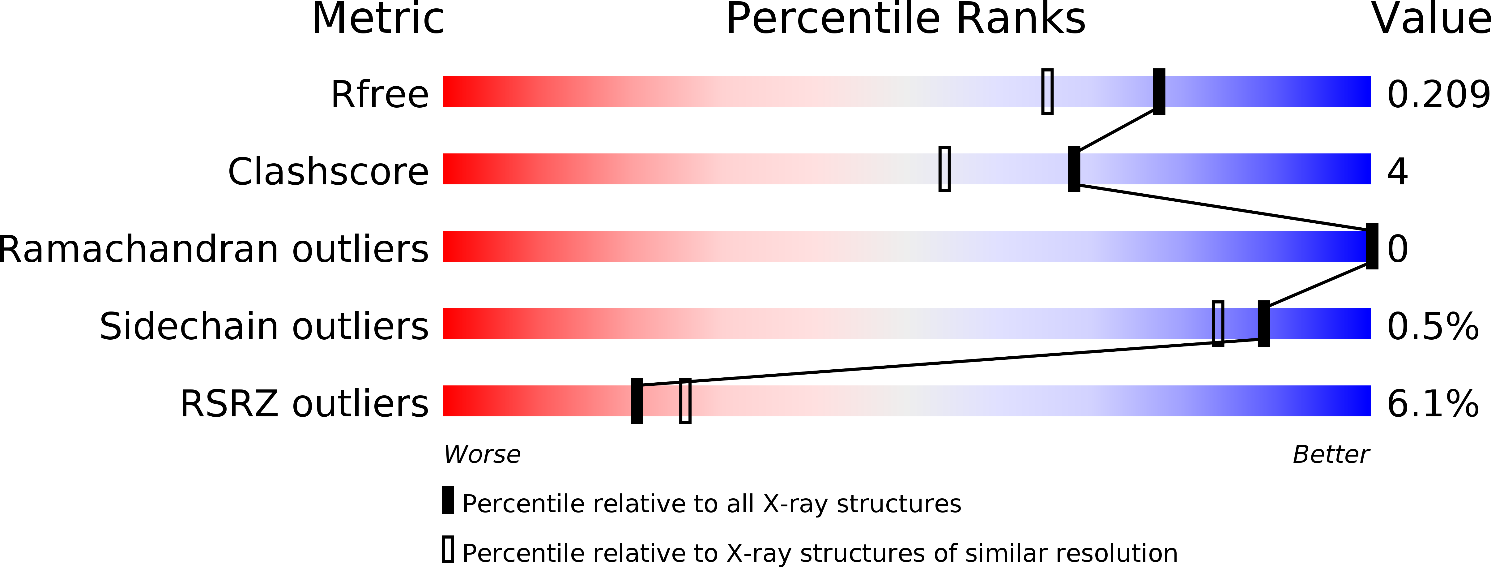
Resolution:
1.76 Å
R-Value Free:
0.20
R-Value Work:
0.16
R-Value Observed:
0.17
Space Group:
C 1 2 1
Macromolecular Entities
Polymer Type:polypeptide(L)
Description:Circadian clock protein kinase KaiC
Chain IDs:A, B, C
Chain Length:253
Number of Molecules:3
Biological Source:Synechococcus elongatus (strain PCC 7942)
Primary Citation
Circadian rhythms. Atomic-scale origins of slowness in the cyanobacterial circadian clock.
Science 349 312 316 (2015)
PMID: 26113637 DOI: 10.1126/science.1261040

Abstact

Circadian clocks generate slow and ordered cellular dynamics but consist of fast-moving bio-macromolecules; consequently, the origins of the overall slowness remain unclear. We identified the adenosine triphosphate (ATP) catalytic region [adenosine triphosphatase (ATPase)] in the amino-terminal half of the clock protein KaiC as the minimal pacemaker that controls the in vivo frequency of the cyanobacterial clock. Crystal structures of the ATPase revealed that the slowness of this ATPase arises from sequestration of a lytic water molecule in an unfavorable position and coupling of ATP hydrolysis to a peptide isomerization with high activation energy. The slow ATPase is coupled with another ATPase catalyzing autodephosphorylation in the carboxyl-terminal half of KaiC, yielding the circadian response frequency of intermolecular interactions with other clock-related proteins that influences the transcription and translation cycle.

Legend

Protein

Chemical

Disease

Primary Citation of related structures