4S2R image
Deposition Date 2015-01-22
Release Date 2015-04-22
Last Version Date 2023-09-20
Entry Detail
PDB ID:
4S2R
Keywords:
Title:
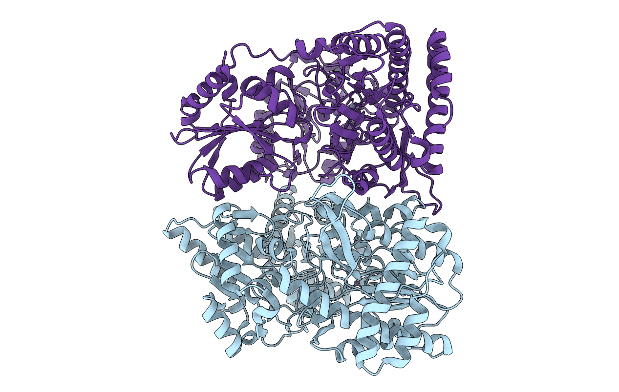
Crystal structure of X-prolyl aminopeptidase from Caenorhabditis elegans: a cytosolic enzyme with a di-nuclear active site
Biological Source:
Source Organism:
Host Organism:
Method Details:
Experimental Method:
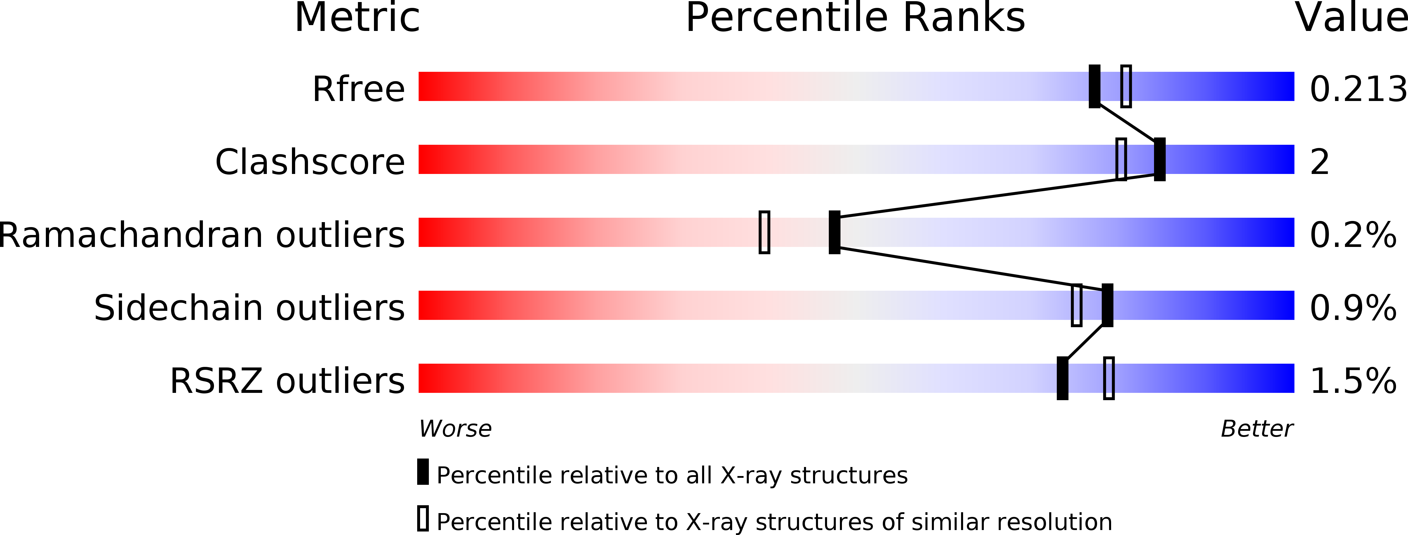
Resolution:
1.95 Å
R-Value Free:
0.21
R-Value Work:
0.16
R-Value Observed:
0.16
Space Group:
C 1 2 1
Macromolecular Entities
Structures with similar UniProt ID
Protein Blast
Polymer Type:polypeptide(L)
Molecule:Protein APP-1
Gene (Uniprot):app-1
Chain IDs:A (auth: P), B (auth: Q)
Chain Length:639
Number of Molecules:2
Biological Source:Caenorhabditis elegans
Ligand Molecules
Primary Citation
Crystal structure of X-prolyl aminopeptidase from Caenorhabditis elegans: A cytosolic enzyme with a di-nuclear active site.
FEBS Open Bio 5 292 302 (2015)
PMID: 25905034 DOI: 10.1016/j.fob.2015.03.013

Abstact

Eukaryotic aminopeptidase P1 (APP1), also known as X-prolyl aminopeptidase (XPNPEP1) in human tissues, is a cytosolic exopeptidase that preferentially removes amino acids from the N-terminus of peptides possessing a penultimate N-terminal proline residue. The enzyme has an important role in the catabolism of proline containing peptides since peptide bonds adjacent to the imino acid proline are resistant to cleavage by most peptidases. We show that recombinant and catalytically active Caenorhabditis elegans APP-1 is a dimer that uses dinuclear zinc at the active site and, for the first time, we provide structural information for a eukaryotic APP-1 in complex with the inhibitor, apstatin. Our analysis reveals that C. elegans APP-1 shares similar mode of substrate binding and a common catalytic mechanism with other known X-prolyl aminopeptidases.

Legend

Protein

Chemical

Disease

Primary Citation of related structures
Feedback Form
Name
Email
Institute
Feedback