4S1K image
Deposition Date 2015-01-14
Release Date 2015-03-25
Last Version Date 2024-02-28
Entry Detail
PDB ID:
4S1K
Keywords:
Title:
Structure of Uranotaenia sapphirina cypovirus (CPV17) polyhedrin at 100 K
Biological Source:
Source Organism:
Method Details:
Experimental Method:
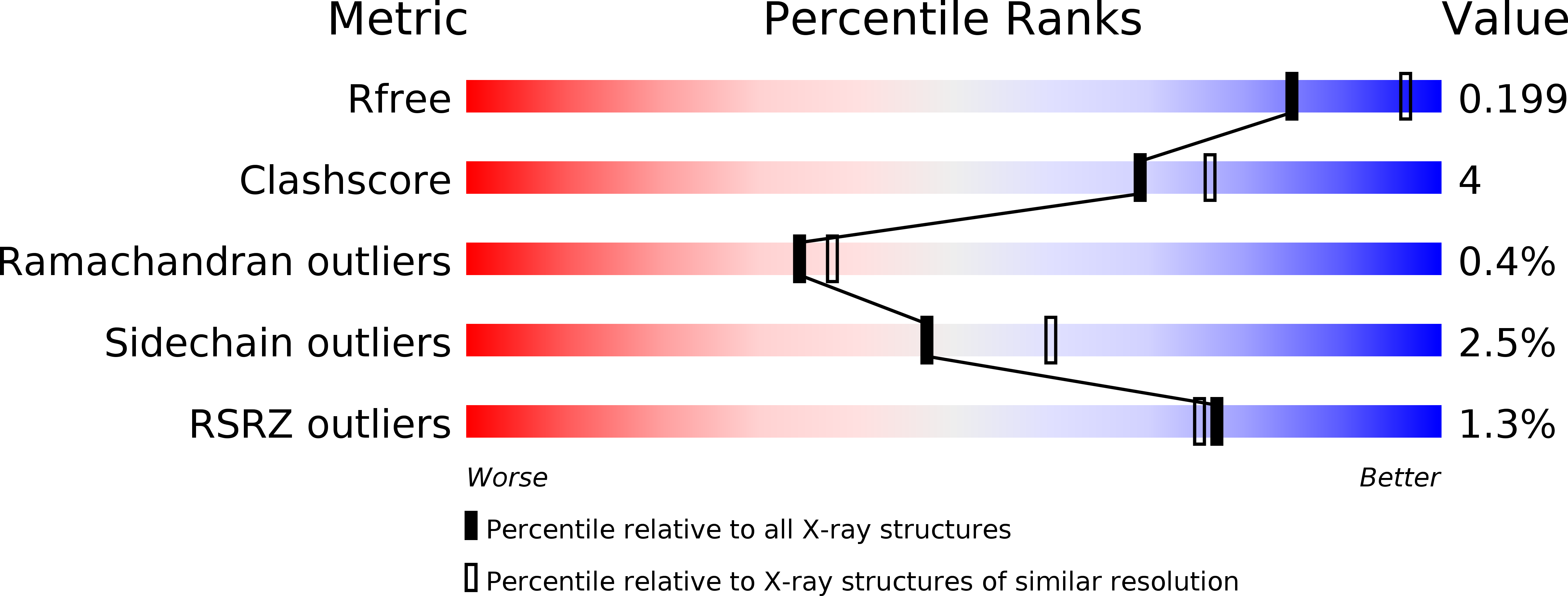
Resolution:
2.20 Å
R-Value Free:
0.19
R-Value Work:
0.14
R-Value Observed:
0.15
Space Group:
I 2 3
Macromolecular Entities
Polymer Type:polypeptide(L)
Molecule:Polyhedrin
Chain IDs:A
Chain Length:237
Number of Molecules:1
Biological Source:Uranotaenia sapphirina cypovirus
Primary Citation
Structure of CPV17 polyhedrin determined by the improved analysis of serial femtosecond crystallographic data.
Nat Commun 6 6435 6435 (2015)
PMID: 25751308 DOI: 10.1038/ncomms7435

Abstact

The X-ray free-electron laser (XFEL) allows the analysis of small weakly diffracting protein crystals, but has required very many crystals to obtain good data. Here we use an XFEL to determine the room temperature atomic structure for the smallest cytoplasmic polyhedrosis virus polyhedra yet characterized, which we failed to solve at a synchrotron. These protein microcrystals, roughly a micron across, accrue within infected cells. We use a new physical model for XFEL diffraction, which better estimates the experimental signal, delivering a high-resolution XFEL structure (1.75 Å), using fewer crystals than previously required for this resolution. The crystal lattice and protein core are conserved compared with a polyhedrin with less than 10% sequence identity. We explain how the conserved biological phenotype, the crystal lattice, is maintained in the face of extreme environmental challenge and massive evolutionary divergence. Our improved methods should open up more challenging biological samples to XFEL analysis.

Legend

Protein

Chemical

Disease

Primary Citation of related structures