4S0P image
Deposition Date 2015-01-04
Release Date 2016-07-20
Last Version Date 2023-09-20
Entry Detail
PDB ID:
4S0P
Keywords:
Title:
Crystal Structure of the Autoinhibited Dimer of Pro-apoptotic BAX (II)
Biological Source:
Source Organism:
Homo sapiens (Taxon ID: 9606)
Host Organism:
Method Details:
Experimental Method:
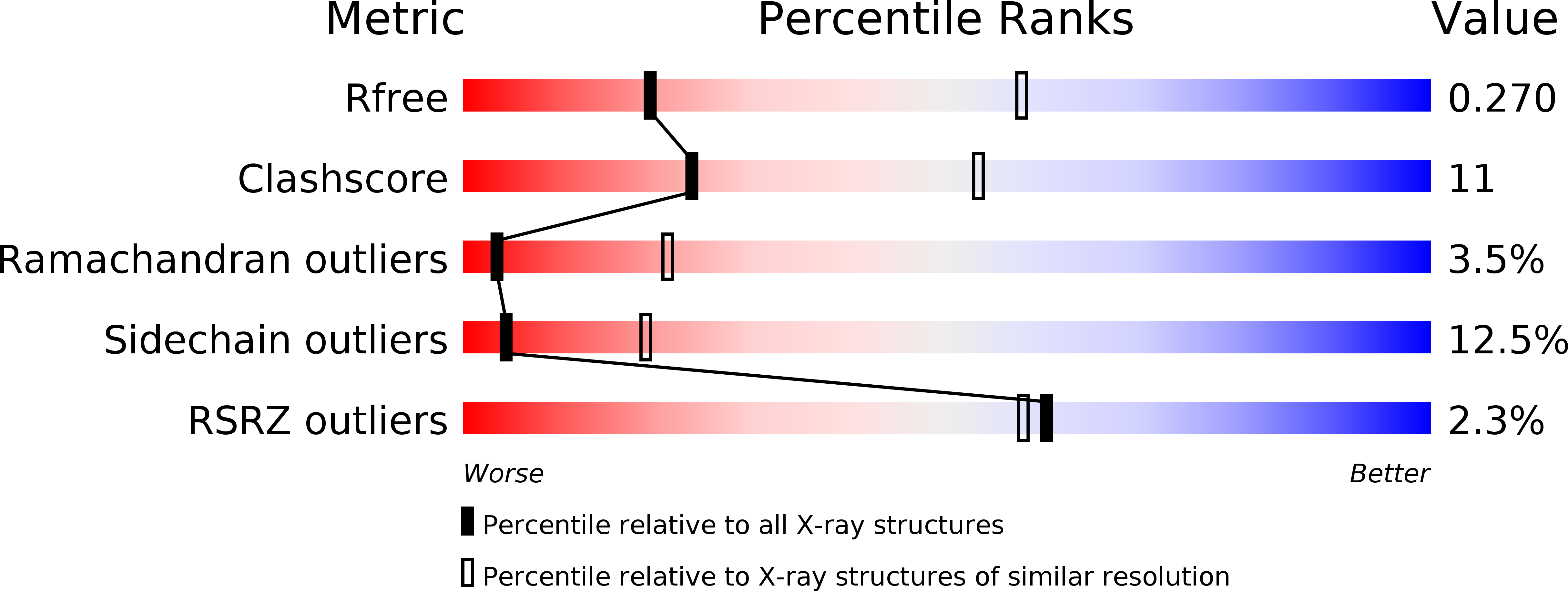
Resolution:
3.25 Å
R-Value Free:
0.26
R-Value Work:
0.21
R-Value Observed:
0.21
Space Group:
P 1 21 1
Macromolecular Entities
Structures with similar UniProt ID
Protein Blast
Polymer Type:polypeptide(L)
Molecule:Apoptosis regulator BAX
Gene (Uniprot):BAX
Mutagens:G67R
Chain IDs:A, B
Chain Length:192
Number of Molecules:2
Biological Source:Homo sapiens
Ligand Molecules
Primary Citation
An Autoinhibited Dimeric Form of BAX Regulates the BAX Activation Pathway.
Mol.Cell 63 485 497 (2016)
PMID: 27425408 DOI: 10.1016/j.molcel.2016.06.010

Abstact

Pro-apoptotic BAX is a cell fate regulator playing an important role in cellular homeostasis and pathological cell death. BAX is predominantly localized in the cytosol, where it has a quiescent monomer conformation. Following a pro-apoptotic trigger, cytosolic BAX is activated and translocates to the mitochondria to initiate mitochondrial dysfunction and apoptosis. Here, cellular, biochemical, and structural data unexpectedly demonstrate that cytosolic BAX also has an inactive dimer conformation that regulates its activation. The full-length crystal structure of the inactive BAX dimer revealed an asymmetric interaction consistent with inhibition of the N-terminal conformational change of one protomer and the displacement of the C-terminal helix α9 of the second protomer. This autoinhibited BAX dimer dissociates to BAX monomers before BAX can be activated. Our data support a model whereby the degree of apoptosis induction is regulated by the conformation of cytosolic BAX and identify an unprecedented mechanism of cytosolic BAX inhibition.

Legend

Protein

Chemical

Disease

Primary Citation of related structures