Deposition Date
2015-01-02
Release Date
2015-05-27
Last Version Date
2025-02-12
Entry Detail
PDB ID:
4S0N
Keywords:
Title:
Crystal Structure of HLTF HIRAN Domain bound to DNA
Biological Source:
Source Organism(s):
Homo sapiens (Taxon ID: 9606)
synthetic construct (Taxon ID: 32630)
synthetic construct (Taxon ID: 32630)
Expression System(s):
Method Details:
Experimental Method:
Resolution:
1.50 Å
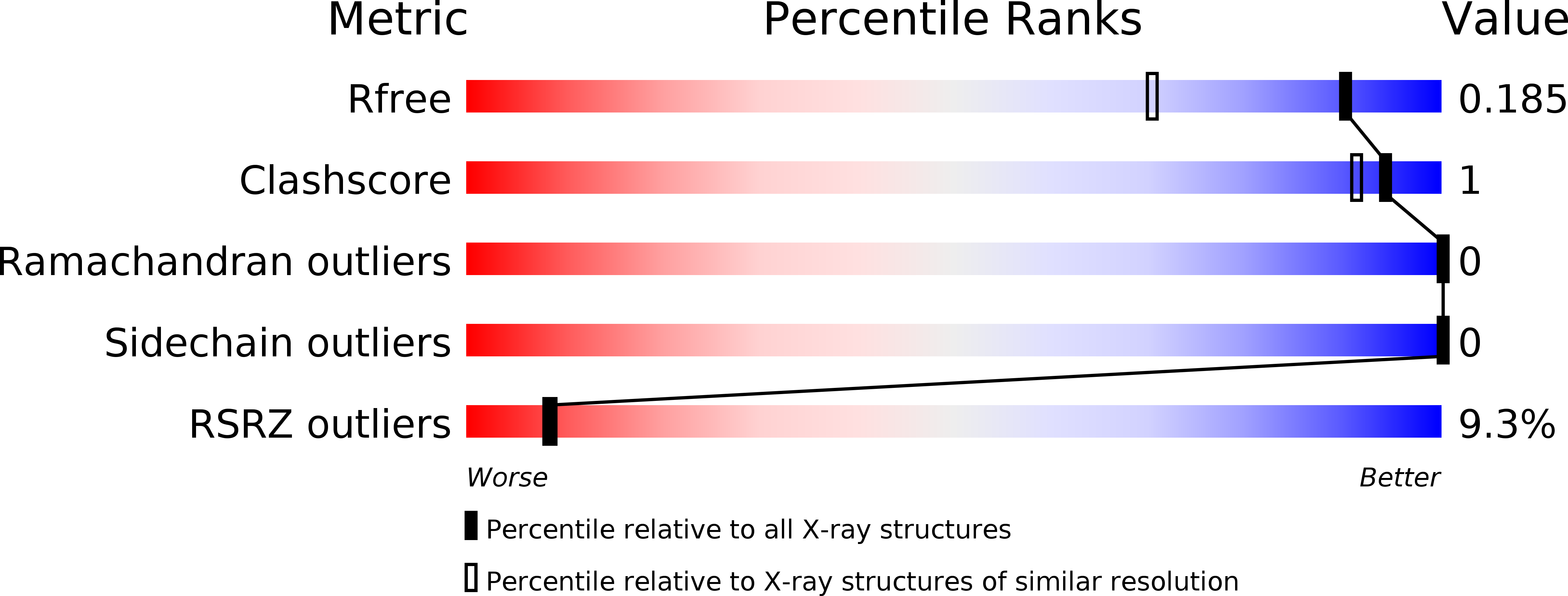
R-Value Free:
0.18
R-Value Work:
0.15
R-Value Observed:
0.15
Space Group:
P 1 21 1