4RWU image
Deposition Date 2014-12-05
Release Date 2014-12-17
Last Version Date 2023-09-20
Entry Detail
PDB ID:
4RWU
Keywords:
Title:
J-domain of Sis1 protein, Hsp40 co-chaperone from Saccharomyces cerevisiae
Biological Source:
Source Organism:
Host Organism:
Method Details:
Experimental Method:
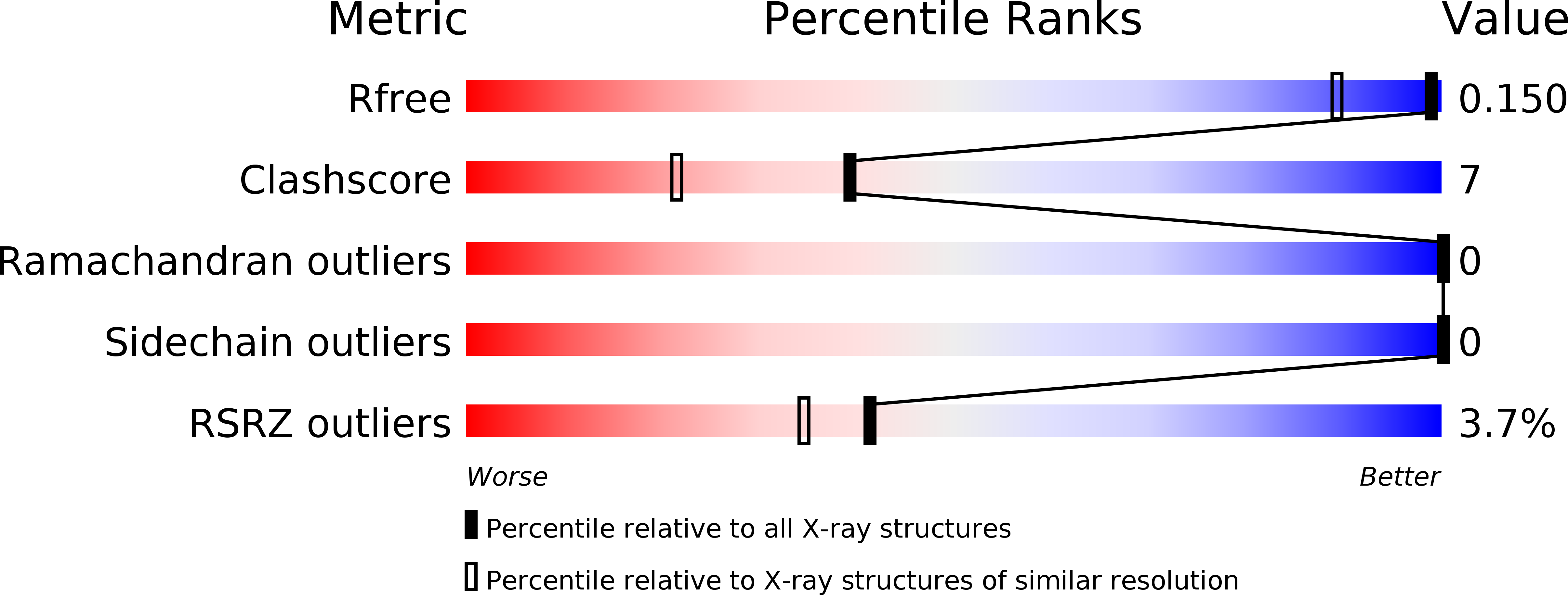
Resolution:
1.25 Å
R-Value Free:
0.14
R-Value Work:
0.12
R-Value Observed:
0.12
Space Group:
P 21 21 21
Macromolecular Entities
Structures with similar UniProt ID
Protein Blast
Polymer Type:polypeptide(L)
Molecule:Protein SIS1
Gene (Uniprot):SIS1
Chain IDs:A
Chain Length:92
Number of Molecules:1
Biological Source:Saccharomyces cerevisiae S288c
Primary Citation
Roles of intramolecular and intermolecular interactions in functional regulation of the Hsp70 J-protein co-chaperone Sis1.
J.Mol.Biol. 427 1632 1643 (2015)
PMID: 25687964 DOI: 10.1016/j.jmb.2015.02.007

Abstact

Unlike other Hsp70 molecular chaperones, those of the eukaryotic cytosol have four residues, EEVD, at their C-termini. EEVD(Hsp70) binds adaptor proteins of the Hsp90 chaperone system and mitochondrial membrane preprotein receptors, thereby facilitating processing of Hsp70-bound clients through protein folding and translocation pathways. Among J-protein co-chaperones functioning in these pathways, Sis1 is unique, as it also binds the EEVD(Hsp70) motif. However, little is known about the role of the Sis1:EEVD(Hsp70) interaction. We found that deletion of EEVD(Hsp70) abolished the ability of Sis1, but not the ubiquitous J-protein Ydj1, to partner with Hsp70 in in vitro protein refolding. Sis1 co-chaperone activity with Hsp70∆EEVD was restored upon substitution of a glutamic acid of the J-domain. Structural analysis revealed that this key glutamic acid, which is not present in Ydj1, forms a salt bridge with an arginine of the immediately adjacent glycine-rich region. Thus, restoration of Sis1 in vitro activity suggests that intramolecular interactions between the J-domain and glycine-rich region control co-chaperone activity, which is optimal only when Sis1 interacts with the EEVD(Hsp70) motif. However, we found that disruption of the Sis1:EEVD(Hsp70) interaction enhances the ability of Sis1 to substitute for Ydj1 in vivo. Our results are consistent with the idea that interaction of Sis1 with EEVD(Hsp70) minimizes transfer of Sis1-bound clients to Hsp70s that are primed for client transfer to folding and translocation pathways by their preassociation with EEVD binding adaptor proteins. These interactions may be one means by which cells triage Ydj1- and Sis1-bound clients to productive and quality control pathways, respectively.

Legend

Protein

Chemical

Disease

Primary Citation of related structures